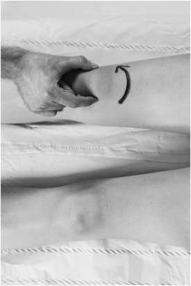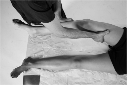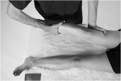
Rationale: Excessive lateral translation of the patella, compared to the contralateral side, may suggest previous dislocation and injury to the medial patellofemoral ligament and retinaculum.
Technique: The patient is supine and the knee is in extension. The examiner holds the medial and lateral patella edges and gently displaces the patella laterally, whilst carefully assessing change in the patient’s facial expressions.
Positive test: Patient apprehension and asymmetrical lateral subluxation of the patella compared with the contralateral side. A useful grading system describes the amount of lateral movement as a percentage of the patella width – i.e. 25%, 50%, 75% or 100% lateral translation.
How is patella tracking assessed?*
Rationale: Maltracking patella is a cause of recurrent dislocations and can cause anterior knee pain.
Technique: Assess the linear movement of the patella during active knee flexion and extension with the patient sitting. Medial movement of the patella during the first 30° of knee flexion is found when the patella engages within the trochlea of the distal femur.
What is Clarke’s test?
Rationale: This tests helps isolate the cause of anterior knee pain.
Technique: The test involves compressing the patella against the trochlea during quadriceps muscle contraction to illicit patellofemoral pain. With the patient supine, place pressure over the patella and ask the patient to contract the quadriceps by extending the straight leg – ‘pushing down’ into the examination couch.
Positive test: Reproduction of the patient’s discomfort. Comparison to the contralateral side is essential, as the test can be uncomfortable in a normal knee.
How are the collateral ligaments of the knee assessed?*

Varus pressure tests the lateral collateral ligament.

Valgus pressure tests the medial collateral ligament.
Technique: Varus stress is applied with the knee in extension and 30° of flexion; opening up the lateral joint line when compared to the contralateral knee indicates injury to the lateral collateral ligament and/or posterior lateral corner. Valgus stress in knee extension and 30° of flexion examines the medial collateral ligament and posteromedial capsule.
Positive test: Asymmetrical joint line gapping with no resistance or ‘end point’ to the examination indicates a complete rupture. Partial gapping with some resistance and (often significant) discomfort felt by the patient indicates a partial collateral ligament injury.
How are meniscal tests performed?*
Rationale: There are numerous available in the literature. They all have poor sensitivity and specificity but all aim to reproduce pain or catching from rotatory movements of the tibia.
Technique: The two commonly used tests are the McMurray’s and Thessaly tests.
McMurray’s starts with the knee flexed and stresses the medial joint line with a varus and external rotation force to reproduce clicking or catching during knee extension. This movement is reversed for the lateral meniscus.
The Thessaly test involves a twisting movement with the patient standing on the symptomatic leg with 20° of knee flexion. Reproduction of pain is reported to have a 95% diagnostic accuracy for meniscal tears.16
Stay updated, free articles. Join our Telegram channel

Full access? Get Clinical Tree


