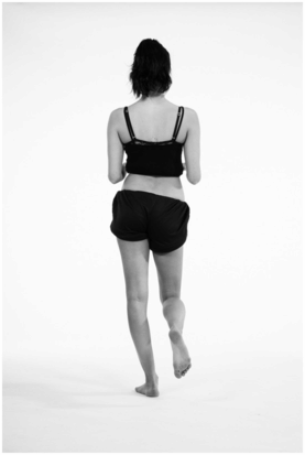
Positive Trendelenburg’s test: weakness of hip abductors on the left side.
Rationale: A Trendelenburg’s test (orthopaedic) identifies weakness of the abductors of the hip.
Technique: The patient is standing facing the examiner. The patient places his or her hands (palms down) on the examiner’s hands (palms up). The patient is asked to ‘stand on one leg’.
Positive test: A contralateral dip of the pelvis or increasing weight placed through the contralateral hand are indicators of weak hip abductors on the standing (ipsilateral) leg. Tilting of the pelvis during walking is known as a Trendelenburg gait.
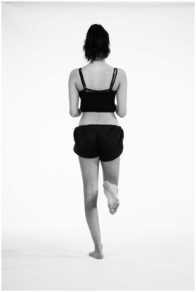
Trendelenburg’s test:
Patient stands on left leg; right leg is flexed at knee and hip.
If the pelvis tilts to the right or the patient presses down with right hand, i.e. ‘the sound side sags’, the abductors on the left side (the standing leg) are weak.
How do you assess for leg length discrepancy?
Apparent leg length is measured from the xiphisternum to the inferior surface of the medial malleolus. Discrepancy in apparent leg length (not true leg length) is caused by a pelvic tilt, scoliosis or hip fixed flexion deformity.
True leg length is measured from the anterior superior iliac spine (ASIS) to the inferior surface of the medial malleolus on the ipsilateral side. Discrepancy in true leg length is a result of shortening of the tibia or femur. If there is a discrepancy in true leg length, Galleazi’s test assesses which bone is involved.
Galleazi’s test is carried out with the patient supine. The heels and knees are placed together, with the knees flexed to 90°. A mismatch in symmetry between thigh and calf length identifies whether the femur or the tibia is short.
What features do you look for during the hip examination?
A systematic approach assessing skin, soft tissues and then bone alignment will help avoid missing pathology.
How do you palpate the hip joint?
The hip joint is a deep joint covered by large muscles and cannot be palpated directly. The soft tissues along the medial side of the thigh can be felt for adductor tenderness. Tenderness over the greater trochanter would suggest bursitis or gluteal tendinopathy, whilst tenderness over the pubic symphysis may indicate osteitis pubis. Pain in the groin (during movement) may indicate pain arising from the femoral head or acetabulum; direct palpation of these structures is usually not possible. Keep in mind other causes of pain in the groin, such as inguinal or femoral hernias, femoral artery aneurysms or femoral vein thrombosis.
How is movement of the hip joint assessed?
The range of movement of the hip joint is described in the table below. The absolute range of movement of the hip is variable. The most important assessment is comparison with the contralateral side.
| Hip movement | Typical range of movement |
|---|---|
| Flexion | 120–140° |
| Extension | 30° |
| Internal rotation | 30–60° |
| External rotation | 30–60° |
| Adduction | 30° |
| Abduction | 45° |
Stay updated, free articles. Join our Telegram channel

Full access? Get Clinical Tree


