Carpal tunnel decompression scar. Radial border of the ring finger and Kaplan’s cardinal line are the key landmarks shown (dotted lines). If the incision needs to be extended proximally past the distal wrist crease, the proximal end should not go radially, but should be extended towards the ulnar side. This is needed to avoid the cutaneous branch of the median nerve, which is found just radial to palmaris longus.

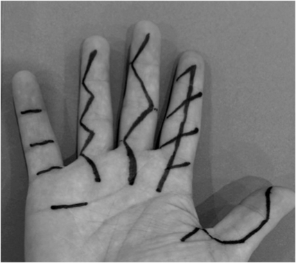
Palmar incisions: thumb, mid-lateral mixed with Bruner; index, Skoog incorporating Z-plasties; middle, Bruner; ring, Littler; little, McCash.
What is the sequence of joints in the hand, from proximal to distal?
Radiocarpal joint
Mid-carpal joint
CMC (carpometacarpal) joint
MCP (metacarpophalangeal) joint
PIP (proximal interphalangeal) joint
DIP (distal interphalangeal) joint
What are the common hand deformities?
Dupuytren’s contractures
Rheumatoid hands
Arthritic: OA, psoriatic or seronegative (SLE) arthritides
Dropped ‘mallet’ finger (extensor tendon rupture)
What are the common finger deformities?
Swan neck: hyperextended PIP joint, flexed DIP joint (common in RA)
Boutonniere: flexed PIP joint, hyperextended DIP joint (common in RA)
Mallet: flexed (unable to actively extend) DIP joint (distal extensor insertion rupture)
Bouchard’s nodes: PIP joint osteophytes in OA
Heberden’s nodes: DIP joint osteophytes in OA
Finger (MCP joint) ulnar deviation in RA
Trigger finger: finger gets stuck in flexion due to tendon nodule stuck under A1 pulley
What are the examination findings in Dupuytren’s disease?
Palmar pits, palmar Dupuytren’s nodules and cords and dorsal knuckle (Garrod’s) pads.
Contraction of the palmar or digital fascia with resulting flexion deformity of MCP and PIP joints, commonly of little and ring fingers.
Pathological cords are: pre-tendinous (palmar), central (digital), lateral (lateral digital), spiral around digital nerve (combination of aforementioned), natatory (web-space).
Hueston’s tabletop test is positive if the patient is unable to place all fingers flat on a table, signifying flexion contractures that merit surgical correction.
What scars can be seen in patients who have had surgery for Dupuytren’s disease?
Longitudinal or transverse (to access > 1 ray) palmar and digital skin incisions. Fasciectomy involves excision of diseased fascia to the affected digit (limited) or all palmar fascia (radical). Skin closure by z-plasty or skin graft.
Skin and fascia excision (dermofasciectomy with FTSG) may reduce recurrence.
What are the examination findings in rheumatoid arthritis?
A symmetrical polyarthropathy (90% hand involvement) with extra-articular manifestations.
Rheumatoid nodules on extensor aspect elbows (prognostic of severe disease).
Joint and tendon synovitis (warm, boggy swelling when active disease).
Wrists: limited extension/supination from caput ulnae syndrome with dorsally prominent ulna head, and palmar subluxed wrist.
Tendons: extensor tendon attrition ruptures over ulnar styloid (Vaughan–Jackson lesion).
Fingers: z-thumb, Boutonnière, swan neck and ulnar drift deformities.
What are the features of osteoarthritis?
Heberden’s and Bouchard’s nodes
‘Squaring’ deformity of the wrists
Crepitus on joint movement
Stiffness, tenderness and reduced ROM
The patient is always seen in the anatomical position: hands opened and supinated, fingers pointing to the floor, palms facing forwards.
Remember that thumb abduction occurs anteriorly, away from the palmar plane. Adduction is the opposite movement, towards the palm.
Extension occurs laterally/radially away from the palm. Flexion is the opposite movement towards the palm.
Opposition is a complex composite movement involving abduction, flexion and circumduction to bring thumb to finger pulps.


