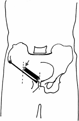
The midpoint of the inguinal ligament is the middle of a line running between the ASIS and the pubic tubercle. This coincides with the deep inguinal ring, which is the origin of indirect inguinal hernias (arrow).

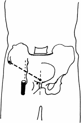
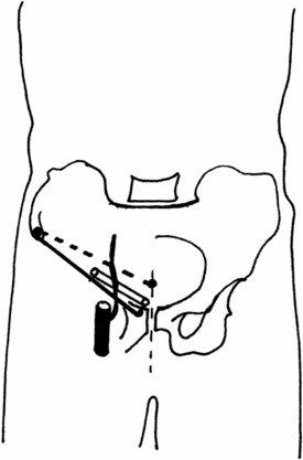
The midinguinal point is the middle of a line running between the ASIS and the public symphisis. This corresponds to the femoral pulse / common femoral artery, and more proximally to the inferior epigastric artery. The inferior epigastric artery arises from the external iliac artery and ascends medial to the deep inguinal ring.

The midpoint of the inguinal ligament is lateral to the midinguinal point. This means that the deep ring of the inguinal canal lies lateral to the inferior epigastric artery. As a result, indirect inguinal hernias are lateral to the inferior epigastric artery and direct ones are found medial to it.
2. Is it a hernia?
First determine if the mass is a hernia. Check if the mass if painful before touching it. Ask the patient to cough to identify a cough impulse, suggestive of a hernia. A pulsatile mass may suggest the presence of a femoral artery aneurysm. A long saphenous vein varix at the saphenofemoral junction may also have a cough impulse, but its location is different from that of a hernia (see later).
3. Is it an inguinal or femoral hernia?
Place two fingers of the right hand on the pubic tubercle and ask the patient to cough.
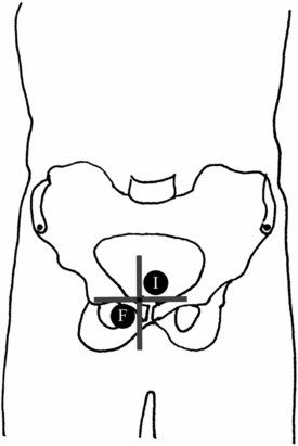
Inguinal hernias travel through the inguinal canal and may emerge through the superficial inguinal ring. An inguinal hernia will thus manifest as a mass above the inguinal ligament and medial to the pubic tubercle (where the inguinal canal terminates as the superficial ring). Inguinal hernias will therefore be superior and medial to the pubic tubercle.
Femoral hernias travel through the femoral canal. This is inferior to the inguinal ligament. Femoral hernias will therefore emerge as a lump inferior and lateral to the pubic tubercle.
4. Is it an inguinal or inguinoscrotal hernia?
Palpate the scrotum to determine if the hernia extends inferiorly into the scrotum. Inguinoscrotal hernias may be more difficult to repair, especially if a laparoscopic approach is used. At this stage also determine if both testes are present, clearly defined and normal. An undescended testis in the inguinal canal may mimic an inguinal hernia. Its absence from the scrotum must therefore be identified. A hydrocoele of the cord may also be confused with an inguinoscrotal hernia. Proceed to a formal scrotal examination if any abnormalities are detected.
Stay updated, free articles. Join our Telegram channel

Full access? Get Clinical Tree


