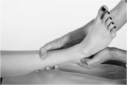
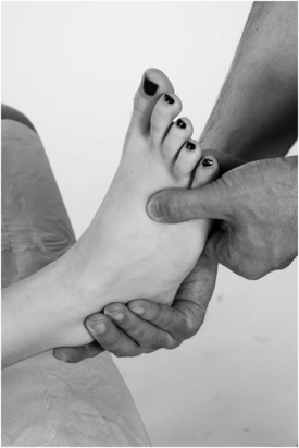
Technique: With the patient sitting and 10° of plantarflexion, stabilise the tibia, with one hand and cup the heel with the other, apply an anterior force to the foot.
Positive test: As with the anterior drawer test in the knee, asymmetrical anterior movement of the hindfoot in relation to the tibia confirms an ATFL injury.
The talar tilt test*

Rationale: this is a lateral ligament stress test and helps identify a deltoid or calcaneofibular ligament injury.
Technique: With the patient sitting with the knee flexed to 90°, a valgus or varus force is applied across the ankle joint, with one hand cupping the heel and the other the tibia.
Positive test:
Asymmetrical opening up in valgus stress indicates a deltoid or medial ligament injury.
Asymmetrical opening up in varus stress indicates a calcaneofibular ligament injury.
Examination for Morton’s neuroma*

Technique: A palpable or audible click, known as a Mulder’s click, felt during compression of the forefoot. Importantly, neuromas are rarely found in the 4th metatarsal space, and are virtually unknown in the first metatarsal space. In these cases careful consideration of differentials is essential.
Positive test: A palpable click, typically felt between the 3rd and 4th metatarsal heads.


