Division of the abdomen into nine regions. RH, right hypochondrium; Ep, epigastric area; LH, left hypochondrium; RL, right lumbar area; Um, umbilical or periumbilical area; LL, left lumbar area; RI, right iliac fossa / right inguinal area; Hg/SP, hypogastric or suprapubic area; LI, left iliac fossa / left inguinal area.
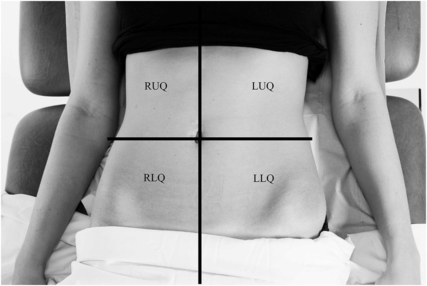
Division of the abdomen into four quadrants. RUQ, right upper quadrant; LUQ, left upper quadrant; RLQ, right lower quadrant; LLQ, left lower quadrant.
What organs can be palpated in each abdominal area?
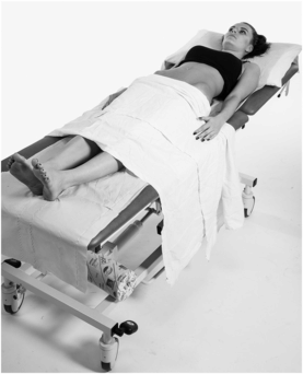
Position and exposure of the patient for abdominal examination. The patient must lie flat with a pillow under the head. Note that the legs and back must be exposed, in addition to the abdomen. The abdomen must be exposed from the lower chest (below the breasts) to the pubis, with access to the groins for examination of the inguinal areas, genitalia and femoral pulses.
When examining the abdomen, the surgeon must ask three questions:
1. Is there any surgical disease or process involving the skin (layer 1)?
2. Is there any abdominal wall pathology (layer 2)?
3. Is there any pathology arising from the abdominal cavity (layer 3)?
Layer 1: What skin changes should be identified?
Scars (previous surgery), stomas, sinuses, fistulas, lacerations
Trauma: haematomas or ecchymosis
Infective: erythema, cellulitis, abscesses
Tumours: benign or malignant growths
Layer 2: What abdominal wall changes should be identified?
Hernias
Soft tissue masses
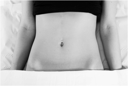
Inspection of the abdomen. This should be done from the right side of the bed, inspecting from above, but also from the side of the abdomen to look for changes in abdominal contour (distension, peristalsis, pulastions). The back and flanks must also be visualised as part of the abdominal inspection.
Where should hernias be identified?
A hernia is ‘an abnormal protrusion of an organ or tissue through the wall of the cavity in which it is contained’. Hernias must be actively sought out and excluded through inspection and palpation in the following areas:
inguinal canal
femoral canal
obturator canal
peri- or intraumbilical area
pararectal space (Spigelian hernia)
midline of the abdomen (epigastrium)
deep to scars (incisional)
Patients can be asked to cough or lift their head or feet off the bed in order to raise intra-abdominal pressure and make hernias apparent.
Any abdominal, flank or groin incision needs to be examined to rule out incisional herniation.
Midline ventral hernias (midline fascial defects) must be differentiated from divarication of the recti (lateral displacement of abdominal recti muscles with stretching of the underlying connective tissues but absence of a fascial defect).
Desmoid tumours are rare causes of pain originating in the abdominal wall. They are fibrous tumours originating in muscular or aponeurotic tissue. Although rare, these are usually found in scar tissue, at the site of previous trauma or in the rectus abdominis muscle. On examination, they are fixed to abdominal wall structures and have normal overlying skin. They occur in Gardner’s syndrome (familial polyposis coli, desmoid tumours, osteomas of the mandible and multiple sebaceous cysts).
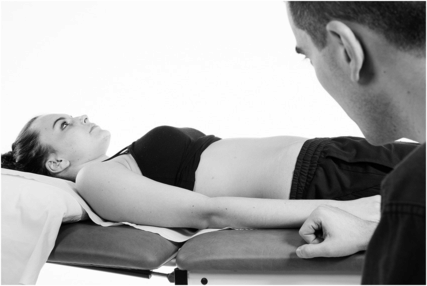
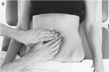
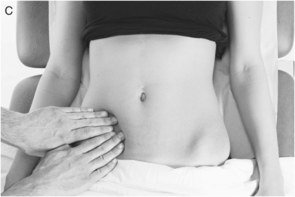
Testing the abdominal wall for hernias: (A) ventral hernias and (B) umbilical or paraumbilical hernias. Hernias are more apparent with increased intra-abdominal pressure. The patient should be asked to cough, and to lift the legs off the bed, and should finally be examined standing.
What is the purpose of light palpation?
Light palpation aims to determine whether there is:
Peritoneal irritation: generalised or localised pain, rigidity or rebound guarding
Distension: 5 Fs (see below)
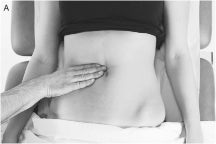
What is the purpose of deep palpation?
Deep palpation investigates tenderness or changes in individual organs in the nine abdominal regions.
Palpation of (A) liver and (B) spleen is done in inspiration, in both cases starting in (C) the right iliac fossa and moving towards the right and left upper quadrants, respectively. Palpation of the appendix starts away from the area of pain and ends in the right lower quadrant (C), where rebound tenderness is tested. (D) The kidney is balloted with two hands.

What are the causes of abdominal distension?
The 5 Fs:
Fat
Faeces (constipation)
Flatus (gas, i.e. obstruction)
Fluid (ascites, haemorrhage)
Fetus (pregnancy; also note other gynaecological causes of distension such as fibroids or large ovarian cysts)
Where can the small and large bowel be palpated?
Large and small bowel can be palpated in any part of the abdomen, as they are mobile, partially fixed structures. In the context of bowel obstruction, the abdomen will be distended and resonant on percussion due to gas-filled bowel loops. Alternatively, the abdomen may be dull on percussion if the bowel loops are filled with intestinal fluid or blood.
How is visceral abdominal pain explained from an embryological perspective?
Epigastric pain: viscera arising from the foregut give rise to midline dull epigastric pain
Periumbilical pain: viscera arising from the midgut give rise to midline dull periumbilical pain
Suprapubic pain: viscera arising from the hindgut give rise to midline dull suprapubic pain
What are the anatomical origins of abdominal pain?
In craniocaudal order:


