Fig. 16.1 Cross-section of the penis, showing structures involved in erection.
This diagram shows only part of the rich nervous and vascular filling and drainage system in the penis. The left-hand area shows the situation in the flaccid penis and the right-hand area the erect penis. The rising pressure during erection limits the venous outflow, thus maintaining the erection. The penis contains three cylinders of erectile tissue: two corpora cavernosa and the corpus spongiosum. The corpus spongiosum contains the urethra. The cylinders of erectile tissue are divided into spaces known as sinusoids or lacunae, which are lined by vascular epithelium. The walls of these spaces are made up of thick bundles of smooth muscle cells within a framework of fibroblasts, collagen and elastin (trabeculae). The erectile tissues are supplied with blood from the cavernosal and helicine arteries, which drain into the sinusoidal spaces. Blood is drained from the sinusoidal spaces through emissary veins. The venules join together to form larger veins that drain into the deep dorsal vein or other veins at different parts of the penis. Arterial and sinusoid dilation is important for erection, while swelling is limited by the inelastic tunica albuginea.
There are also many locally produced mediators that appear to be involved in achieving and maintaining an erection. Nitric oxide (NO) synthesised by blood vessel endothelial cells and released from non-adrenergic non-cholinergic (NANC) nerves in the corpora appears to be crucial for cavernosal smooth muscle relaxation. Nitric oxide generates intracellular cGMP, which activates protein kinase G. cGMP is degraded to GMP by phosphodiesterase type 5 (PDE5) (see Table 1.1), terminating its effects. Inhibition of PDE5 is a primary target for the pharmacological treatment of erectile dysfunction (see below). Other vasodilators that are involved in modulating penile vascular smooth muscle relaxation and blood flow include vasoactive intestinal peptide (VIP), calcitonin gene-related peptide (CGRP) and prostaglandin E1, but their precise roles are less well understood. Numerous central facilitatory mediators have been identified, including dopamine, acetylcholine and a variety of peptides. These are involved in the psychological preparedness that is essential for an erection to occur.
Erectile dysfunction
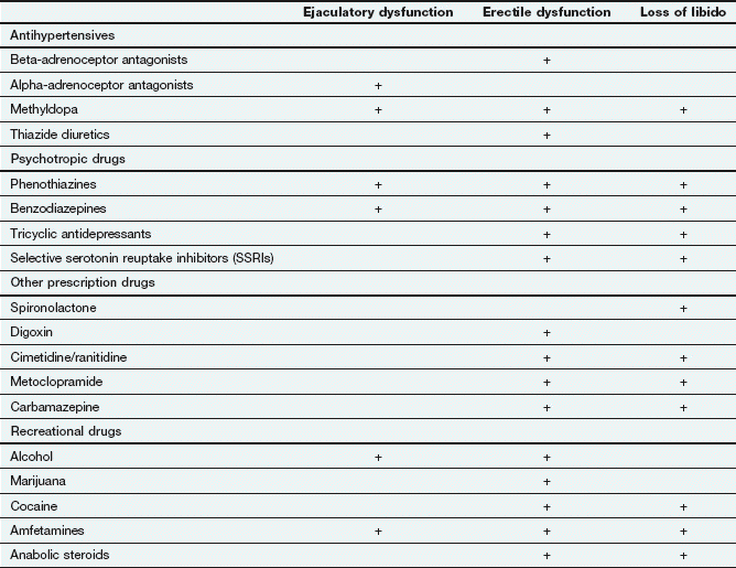
Erectile dysfunction is defined as the consistent inability to achieve or sustain an erection of sufficient rigidity for sexual intercourse. It is a common problem, affecting up to 20% of adult men, with up to 10% over the age of 40 years having complete erectile dysfunction. Any disease process that affects penile neural supply, arterial inflow or venous outflow can produce erectile dysfunction. There is a physical cause in about 80% of cases (Box 16.1) but a psychological component often coexists. Psychogenic erectile dysfunction is more common in younger men. Drugs are an important cause of erectile dysfunction, particularly antihypertensive, psychotropic and ‘recreational’ drugs, and account for up to 25% of cases. Drugs can also affect libido and therefore arousal, or inhibit ejaculation in those who achieve an erection (Table 16.1).
Box 16.1 Common causes of erectile dysfunction
Drugs (see Table 16.1)
Substance abuse, e.g. nicotine, alcohol, recreational drugs
Testosterone deficiency, e.g. hyper- or hypothyroidism
Neurological disease, e.g. multiple sclerosis, Alzheimer’s disease, epilepsy
Psychological factors (20% as a primary cause, more commonly secondary to physical problems)
Management of erectile dysfunction
A number of strategies can be used in the management of erectile dysfunction. Initially there should be an assessment and treatment of any underlying psychological cause or physical disease or if possible withdrawal of a causative drug. Treatment options for persistent dysfunction include:
 mechanical aids, such as the vacuum constriction device: these are usually advised for older people who do not respond to pharmacological treatment and do not wish to have surgery,
Hyperprolactinaemia impairs erection; it is most commonly caused by drug therapy (e.g. with phenothiazines) and can be improved by oral dopamine agonists (Ch. 43) if the cause cannot be treated.
Oral phosphodiesterase inhibitors
Endothelial-derived nitric oxide increases the synthesis of cGMP that acts via protein kinases to cause blood vessel dilation (see Ch. 5, Fig. 5.3). cGMP is broken down in penile tissue by PDE5. Sildenafil, tadalafil and vardenafil are orally active analogues of cGMP that selectively inhibit the enzyme. PDE5 is also found in lower concentrations in other vascular and visceral smooth muscles, and in skeletal muscle and platelets. Sexual stimulation resulting in the release of nitric oxide is a prerequisite for these drugs to produce an erection, and the drug will then prolong the vasodilator effect of nitric oxide on penile vascular smooth muscle. If an appropriate dose of the drug is used, about 60% of men with erectile dysfunction will achieve erections sufficient to permit intercourse. The response is often better if precipitating factors are also treated, such as depression or excess alcohol consumption.
Sildenafil and tadalafil are also used to treat pulmonary hypertension (Ch. 6).
Pharmacokinetics
Sildenafil is relatively well absorbed orally, but vardenafil and tadalafil are less well absorbed. The median time to onset of action for all is about 30 min. The absorption of sildenafil and vardenafil is delayed by a fatty meal, whereas the absorption of tadalafil is rapid and unaffected by food. All are eliminated by hepatic metabolism mediated primarily by CYP3A4. Sildenafil and vardenafil have half-lives of less than 6 h, and should be taken 30–60 min before sexual activity for maximum benefit. The half-life of tadalafil is longer (17 h), and its duration of action is up to about 24–36 h; therefore, planning of sexual activity (and its timing in relation to drug dosage) is less relevant with this drug. Sildenafil and vardenafil both have active metabolites, but tadalafil does not.
Stay updated, free articles. Join our Telegram channel

Full access? Get Clinical Tree


