Epithelioid Hemangioendothelioma
Cyril Fisher, MD, DSc, FRCPath
Key Facts
Terminology
Vascular neoplasm with metastatic potential composed of epithelioid endothelial cells
Clinical Issues
Rare vascular tumor
Superficial or deep soft tissue
Rare in skin
˜ 50% associated with preexisting vessel
Behavior intermediate between hemangioma and angiosarcoma
Metastatic rate (20-30%)
Mortality (10-20%)
Painful mass
All age groups
Wide local excision with clear margins
Adverse prognostic factors
> 3 mitoses per 50 high-power fields
Tumor size > 3 cm
Macroscopic Features
Well-circumscribed nodular lesion
Microscopic Pathology
Rare obvious vascular channels
Short strands, cords, solid nests, or single cells
Bland, epithelioid, round, or slightly spindled endothelial cells
Intracytoplasmic lumina
Can contain red blood cells
Myxohyaline, chondroid-like stroma
Expression of endothelial markers
CD31, CD34, FLI-1
Some cases are cytokeratin positive
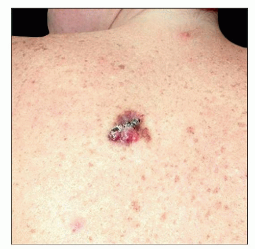 This clinical photograph shows an unusual example of cutaneous epithelioid hemangioendothelioma presenting as a single, discolored, exophytic lesion on the upper back in an adult. |
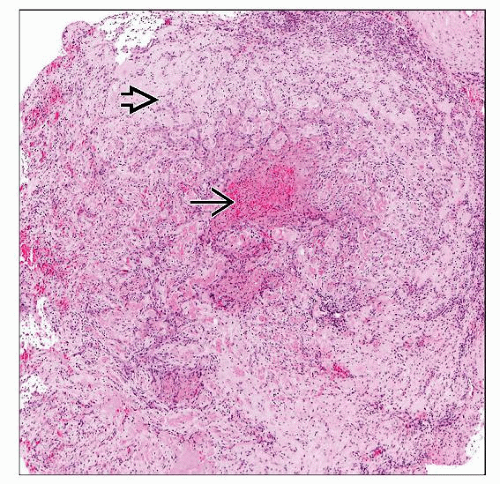 Low-magnification view shows epithelioid hemangioendothelioma with cords of cells  infiltrating the vessel wall and extending into adjacent tissues. The lumen infiltrating the vessel wall and extending into adjacent tissues. The lumen  is filled with thrombotic debris. is filled with thrombotic debris. |
TERMINOLOGY
Abbreviations
Epithelioid hemangioendothelioma (EHE)
Definitions
Angiocentric vascular neoplasm with metastatic potential, composed of epithelioid endothelial cells
CLINICAL ISSUES
Epidemiology
Incidence
Rare vascular tumor
Age
All age groups, but rare in children
Gender
M = F
Site
Skin (rare), superficial or deep soft tissue
Extremities, head and neck, viscera (often multicentric)
Presentation
Painful mass
Solitary mass
Multicentric in a number of cases
Edema in some cases
Occlusion of vessels
Due to tumor origin in/association with preexisting vessels
Can result in ischemic or venous obstructive symptoms
Treatment
Surgical approaches
Wide local excision with clear margins
Prognosis
Behavior intermediate between hemangioma and angiosarcoma
Local recurrence rate 10-15%
Metastatic rate 20-30%, mortality 10-20%
Superficial cases have better prognosis
Adverse prognostic factors
> 3 mitoses per 50 high-power fields
Tumor size > 3 cm
MACROSCOPIC FEATURES
General Features
Well-circumscribed nodular lesion
Intravascular mass resembling organizing thrombus
Stay updated, free articles. Join our Telegram channel

Full access? Get Clinical Tree


