Emphysema
Timothy C. Allen
Philip T. Cagle
Emphysema is characterized by destruction of respiratory bronchioles, alveolar ducts, and alveoli leaving enlarged airspaces. There are several types of emphysema, but the most common type is centrilobular (also known as centriacinar or proximal acinar) emphysema caused by tobacco smoking that involves primarily the respiratory bronchioles. It is most prominent in the upper lung zones. Panacinar emphysema, like that seen with α1-antitrypsin deficiency, involves alveolar ducts as well as the respiratory bronchioles.
The destruction of lung parenchyma in emphysema leaves residual fragments of tissue, including islands of fibrovascular tissue. Although it has been emphasized that emphysema is not the same as diffuse interstitial fibrosis, remodeling of lung tissue also appears to have a role in emphysema, and there may be mild fibrosis. Additional histopathologic changes related to tobacco smoking, including respiratory and membranous bronchiolitis, respiratory bronchiolitis-associated interstitial lung disease, and desquamative and related findings, may be seen in association with emphysema from tobacco smoking. Anthracotic pigment and/or macrophages containing smoker’s pigment may be present. Bullae are cystic spaces that are found in the lung apices, subpleurally, and elsewhere in emphysematous lungs.
Histologic Features
Fragmentation and loss of respiratory bronchioles and septal tissue creating enlarged spaces.
Bullae are typically subpleural and have residual islands of fibrovascular tissue within grossly evident enlarged spaces.
Other findings related to tobacco smoking may be observed, including anthracotic pigment, smoker’s pigment, respiratory bronchiolitis, membranous bronchiolitis, respiratory bronchiolitis-associated interstitial lung disease, and related conditions.
Tissue sections may show mild fibrosis and secondary changes, including focal scars and focal acute or organizing pneumonia.
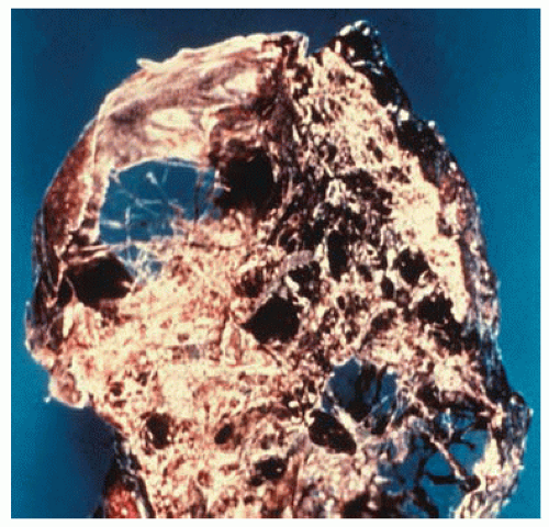 Figure 77.1 Lung with severe emphysema showing extensive destruction of lung parenchyma. |
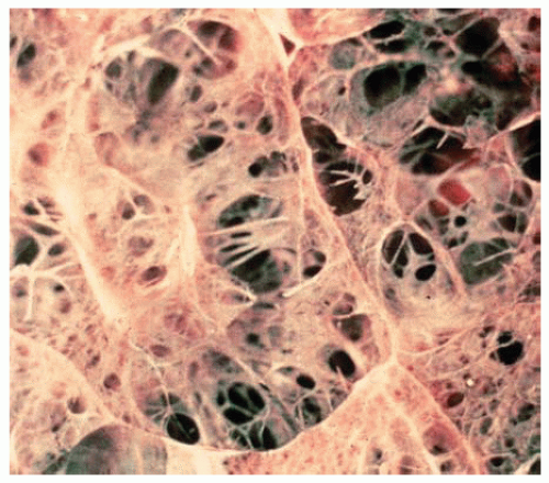 Figure 77.2 Residual strands of fibrovascular tissue in severe emphysema cross-spaces created by destruction of lung parenchyma.
Stay updated, free articles. Join our Telegram channel
Full access? Get Clinical Tree
 Get Clinical Tree app for offline access

|