Embryology and Developmental Lesions


Embryology and Developmental Lesions


Paul W. Biddinger


EMBRYOLOGY


Embryologic Origination

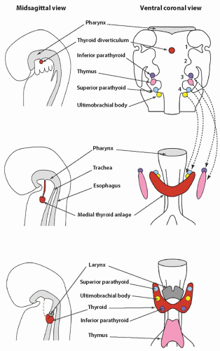
Knowledge of the embryologic development of the thyroid is key to understanding several abnormalities of the thyroid gland. The gland originates from the embryonic foregut, the same structure from which the pharynx, lungs, and upper digestive tract develop. The gland eventually contains two different types of hormonally active cells, follicular and C cells, and each type develops from a different embryologic structure. Follicular cells, which constitute the largest cell population, derive from the medial thyroid anlage. The C cells migrate to join the medial anlage through the lateral anlagen, also known as the ultimobranchial bodies (Fig. 2.1).


Medial Thyroid Primordium

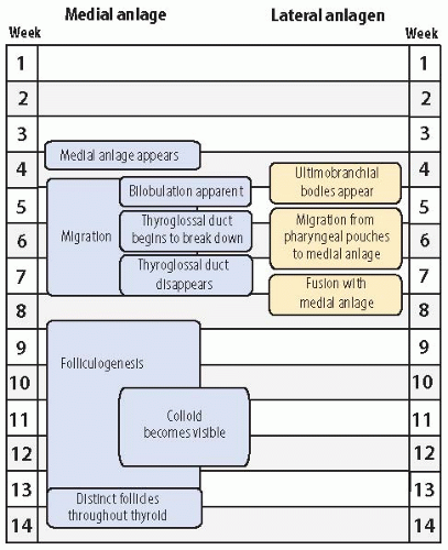
The medial primordium, or anlage, is first visible near the end of the third week of embryonic life (Fig. 2.2). It appears as a thickening of endodermal epithelium of the cranial portion of the foregut, also known as the pharyngeal gut or pharynx.1 The epithelial thickening develops in the ventral midline of the embryonic pharynx between the tuberculum impar and the copula (or hypobranchial eminence). This site, at the level of the second pharyngeal (or branchial) arch, eventually becomes the foramen cecum. The medial primordium soon invaginates to form a pit.


Migration

The invagination becomes more pronounced, yielding a flasklike diverticulum that begins to migrate caudally by the middle of the fourth week, concomitant with the descent of the heart. The migration of the thyroid anlage occurs through the loose mesenchyme ventral to the foregut. The cells continue to proliferate, and bilobation is evident early in the fifth week. The anlage, initially a hollow structure, solidifies with cells. It remains connected to the floor of the pharynx by the thyroglossal duct until the latter part of the fifth week when the duct begins to break down. The duct generally disappears between the sixth and eighth week, but portions may persist as fibrous cords or fine tubular structures with epithelial lining. Persistence of the latter into postnatal life can be the nidus of a thyroglossal duct cyst. As the medial anlage migrates, two lobes joined by an isthmus become visible during the sixth week. It reaches its final position anterior to the proximal region of the trachea during the seventh week of gestation.


Folliculogenesis

Formation of follicles begins in the 8th to 10th week as the thyroid transforms from solid cords of cells into rounded aggregates with small central lumina. Colloid is initially seen between the 10th and 12th week, and thyroid hormone is detectable in the fetal serum by the 11th to 12th week.2,3,4 After the 13th week, the follicular cells appear morphologically well developed at the ultrastructural level,4 and by the 14th week, distinct follicles are seen throughout the thyroid.


Growth and Maturation

The weight of the thyroid gland increases in a nearly linear manner from about 5 to 20 mg to 250 to 500 mg between the 10th and 20th week of gestation. The height of follicular cells stays between 12 to and 13 µm during the 10th to 20th week.5 During this period of active folliculogenesis, the number of follicles per unit area initially increases and then declines, reflecting an increase in follicle size and colloid content. The follicular epithelium to colloid ratio declines until the 17th to 18th week. This ratio then remains fairly constant up to about the 29th week, suggesting the attainment of structural maturity early in the second trimester.5,
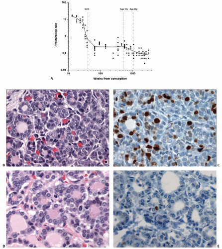
6 Proliferative activity in the thyroid as measured by Ki-67 (MIB-1) immunostaining of nuclei is relatively high between the 10th and 20th week, with averages ranging between 12% and 16% (Fig. 2.3).7 The rate diminishes throughout the latter half of gestation, dropping to <0.5% at the time of birth. The average proliferative rate remains a fraction of a percentage throughout the pediatric and adult periods of life.

The thyroid increases in size throughout gestation, showing the highest relative rate of growth during the second trimester.8 During the second trimester, the gland weight increases 7- to 8-fold, increasing from approximately 100 to 125 mg to 700 to 800 mg.5,6,9 Follicular cell height increases to reach a maximum of 18 µm at about 30 weeks.5 After the 29th week, fetal thyroids tend to show a gradual increase in the epithelial:colloid ratio as epithelial growth outpaces colloid accumulation. The size of follicles decreases and the number of follicles per unit area increases, reflective of the relatively small volume of colloid.

Thyroid glands of newborn full-term infants have a variable appearance in terms of colloid content. A significant number shows no appreciable colloid, and some consider this a physiologic response to labor.10 Desquamation of follicular epithelium and aggregates of pyknotic nuclei can be seen, and some have attributed this to physiologic changes.10 However, in most cases, these latter changes probably represent postmortem autolysis. At end of 40 weeks of gestation, the thyroid gland attains a weight in the range of 1 to 4 g.5,9


Lateral Thyroid Primordia

The ultimobranchial bodies constitute the lateral thyroid primordia that fuse with the medial anlage to form the complete thyroid gland (Fig. 2.1). The ultimobranchial bodies populate the thyroid gland with C cells.11 The precursors of the C cells have been considered derivatives of the neural crest. The neural crest is a transient tissue initially found at the junction of the neural groove and ectoderm. After the neural groove fuses to form the neural tube, the neural crest forms an intermediate zone between the surface ectoderm and neural tube.
The pleuripotent cells of the neural crest migrate extensively throughout the body and differentiate into a wide range of cells including sensory neurons, postganglionic autonomic neurons and Schwann cells of the peripheral nervous system, melanocytes, chondrocytes, and catecholamine-secreting cells of the adrenal medulla.12


FIGURE 2.1. Embryonic thyroid development from the fourth week through the seventh to eighth week. The midsagittal images on the left side show the origin of the medial anlage from the pharynx and the migration to final site adjacent to the larynx and trachea. The ventral coronal view images on the right side show the origin and migration of the inferior parathyroids and thymus (from the third pharyngeal pouches) and the superior parathyroids and ultimobranchial bodies (from the fourth pouches) to their final locations.


FIGURE 2.2. Timeline of thyroid developmental events during embryonic life.

Using chick-quail chimeras, Le Douarin and Le Lièvre demonstrated that avian C cells ultimately derive from the neural crest.13 They engrafted sections of quail neural primordium into chick embryos and traced the migration of the quail cells, distinguishable by their large nucleoli. Quail cells subsequently proved to be the predominant component of the ultimobranchial bodies and formed the C cells.

The assumption that mammalian C cells derive from the neural crest has been challenged recently. The ultimobranchial bodies of birds and lower vertebrates do not fuse with the medial anlage. Avian ultimobranchial bodies are innervated structures containing C cells separate from the thyroid gland, in contrast to mammalian ultimobranchial bodies, which fuse with the medial thyroid and disperse C cells within the gland.14 It is of note that a recent study of mice found that neural crest cells populate pharyngeal arches but not the pouches including the site of the ultimobranchial bodies.15 These findings suggest the origin of murine C cells from the endodermal epithelium as opposed to the neural crest. However, at this time, the orthodox view remains that C cells ultimately derive from the neural crest.

The ultimobranchial bodies become apparent during the fourth to fifth week of gestation as stratified endodermal tissue in contact with the embryonic pharyngeal space.16 The ultimobranchial bodies are located in the most caudal pharyngeal pouches. Because the fifth pouch is rudimentary in humans, controversy exists whether it should be considered a separate fifth pouch or a component of the fourth pharyngeal pouch. Thus, the region containing the ultimobranchial body is sometimes referred to as the fourth-fifth branchial pouch complex.16 For practical purposes in this chapter, ultimobranchial bodies will be considered part of the fourth pouch, acknowledging that they arguably constitute separate fifth pouches.


Migration and Fusion

The ultimobranchial bodies separate from the pharyngeal pouches and migrate centrally to fuse with the medial thyroid usually by the seventh to eighth week of gestation. Fusion typically occurs in the middle or midsuperior regions of the lateral lobes. After fusion with the larger medial anlage, the ultimobranchial bodies undergo dissolution with dispersal of C cells into the surrounding follicular tissue. Portions of the ultimobranchial bodies may persist in fetal or postnatal thyroid glands as small cystic structures or solid cell nests.16,17,18,19


GENES INVOLVED IN THYROID DEVELOPMENT


Early Development

Several genes that appear to play critical roles in thyroid development have been identified (Table 2.1).20,21 The signal that initiates the development of the medial anlage is unknown. However, TTF1 (NKX2-1), TTF2 (FOXE1), PAX8, and HHEX are genes that are involved in the early embryologic stages of thyroid development by cell-autonomous mechanisms. TBX1 has been identified as a gene that also plays an important role in early thyroid development, but, by a cell-nonautonomous mechanism involving the surrounding mesoderm. Much of our knowledge about their roles derives from studies of transgenic mice lacking these genes, the so-called knockout mice.


TTF1

TTF1, also known as NKX2-1, TITF1, or T/EBP, codes for a homeodomain-containing transcription factor, thyroid transcription factor 1 (TTF1), that is able to bind to the promoters of the thyroglobulin gene (Tg) and the thyroperoxidase gene (TPO). It maps to chromosome 14q13 and is a member of the NKX2 family of transcription factors.22 Its expression is coincidental with the appearance of the initial endodermal thickening, and the medial anlage is the only location in the embryonic pharynx that shows TTF1 expression. It remains expressed in the thyroid throughout embryonic and fetal development and into adulthood. TTF1 is also expressed in C cells.23 TTF1 seems to be necessary for the survival of the follicular precursors. Knockout mice lacking this gene show loss of the medial thyroid anlage through apoptosis along with the absence of C cells.24,25 TTF1 does not appear to be necessary for formation and migration of ultimobranchial bodies but is essential for the survival of its cells during migration and for successful fusion with the medial anlage.26

NKX2-5 is another homeobox-containing gene that expresses its protein in the early medial thyroid anlage. NKX2-5 is a transcription factor that has a major role in cardiac development,27 and recent data suggest that it may also play a role in cases of thyroid dysgenesis.28


PAX8

The PAX8 gene codes for a member of a family of transcription factors with a 128 amino acid paired binding domain.29,30 It is located on chromosome 2q12, and it derives its name from paired box gene 8. This transcription factor seems to have synergistic action with TTF1 and is critical for the development of the medial anlage past the early invagination stage.31 Like TTF1, PAX8
appears to be necessary for survival of the anlage but does not initiate its development. PAX8 also appears to play an important role in follicular differentiation. Disruption of both PAX8 alleles in mice results in severe congenital hypothyroidism because of the absence of follicular cells.32 Monoallelic mutations in PAX8 have been documented in patients with sporadic and familial thyroid hypoplasia or ectopy.33,34


FIGURE 2.3. A: Change in the proliferative rate of human thyroid cells by weeks from conception based on immunostaining for Ki-67 (MIB-1), a marker of active cell proliferative cycle. (From Saad AG, Kumar S, Ron E, et al. Proliferative activity of human thyroid cells in various age groups and its correlation with the risk of thyroid cancer after radiation exposure. J Clin Endocrinol Metab. 2006;91:2672-2677, with permission. Copyright 2006, The Endocrine Society.) B-E: Comparison of thyroid of 13-week fetus (B and C) with that of a full-term infant (D and E). (B) and (D) are photomicrographs of hematoxylin and eosin-stained sections, whereas (C) and (E) show immunostaining for Ki-67. The 13-week fetal thyroid shows staining of 16% of nuclei, whereas <1% of nuclei of full-term infant thyroid express Ki-67.


Table 2.1 Genes Involved in Thyroid Development


Gene


Chromosome Location


Functions


TTF1 (NKX2-1)


14q13


• Transcription factor able to bind to promoters of Tg and TPO


• Necessary for survival of follicular precursors


• Necessary for survival of C cells


PAX8


2q12


• Transcription factor having synergistic action with TTF1


• Necessary for survival of medial anlage


• Important for follicular development


TTF2 (FOXE1)


9q22


• Transcription factor able to bind to promoters of Tg and TPO


• Essential role in medial anlage migration


HHEX


10q23


• Unclear; possible maintenance of expression of TTF1, PAX8, and TTF2


TBX1


22q11


• Transcription factor with nonautonomous mechanism involving the surrounding mesoderm


• Appears to regulate the size of early medial anlage through control of FGF8


TSHR


14q31


Codes for thyroid-stimulating hormone receptor


Tg


8q24


• Codes for thyroglobulin


TPO


2p25


• Codes for thyroperoxidase


NIS


19p13


• Codes for sodium-iodide symporter


HOXA3


7p15


• Transcription factor with essential role in the development of pharyngeal glandular tissues


• Important for the development and/or interaction of medial anlage and ultimobranchial bodies


HASH1


12q22-23


• Transcription factor important in the development of C cells


• Important role in the differentiation of neurons derived from neural crest


TTF2

The gene TTF2, also known as FOXE1, TITF2, or FKHL15, codes for a member of the forkhead/winged helix family of transcription factors and maps to chromosome 9q22.35 This transcription factor is a nuclear protein that binds to Tg and TPO promoters. It is expressed when the medial thyroid anlage first appears. Like TTF1 and PAX8, expression continues throughout development and into adulthood. However, it has a wider area of foregut expression, being detected in the developing thyroid, tongue, epiglottis, palate, and esophagus. TTF2 seems to play an essential role in the migration of the medial anlage.


HHEX

The gene HHEX codes for a homeodomain-containing transcription factor that is expressed in the foregut before and after the appearance of the thyroid anlage. The gene is located on chromosome 10q23, and its name derives from hematopoietically expressed homeobox because it was first identified in hematopoietic tissue.36,37 Expression of HHEX is seen in the primordial tissue of thyroid, liver, thymus, pancreas, and lungs. It shows the highest degree of expression in developing and adult thyroid. The role of HHEX is not defined at this time. In particular, it is unclear whether the factor is necessary for maintaining expression of TTF1, TTF2, PAX8, or vice versa.20


TBX1

The gene TBX1, also known as T-box 1, is a member of the family of T-box genes that code for transcription factors involved in the regulation of developmental processes. The gene is located on chromosome 22q11.38 TBX1 plays an important role in the development of pharyngeal structures and is a major gene associated with DiGeorge syndrome.39,40 TBX1 is not expressed in the thyroid primordium but instead is expressed in the surrounding mesoderm, and it appears to regulate the size of the early thyroid primordium through the control of the fibroblast growth factor 8 gene (FGF8) expression.40


Later Development

Genes involved in the later stages of thyroid development include TSHR, Tg, TPO, NIS, HOXA3, FGFR2, and others from the NKX2 family.20 TSHR maps to chromosome 14q31 and is one of the superfamilies of G protein-coupled receptors.41 TSHR, or thyroid-stimulating hormone receptor, is initially expressed in the medial thyroid anlage after the completion of migration but before follicular development. It seems to be important for follicular development because knockout mice with null or loss of function show severe hypothyroidism. These mice have hypoplastic thyroid glands as adults, even though the glands are of normal size at birth.


Tg, TPO, and NIS

Thyroglobulin (Tg), thyroperoxidase (TPO), and sodium-iodide symporter (NIS) are three critical genes involved in thyroid hormone production. Expression of these genes begins after the completion of thyroid anlage migration and continues through prenatal development and throughout postnatal life. Soon after initial expression in the embryonic thyroid, primitive follicles begin to appear. Tg maps to chromosome 8q24 and codes for
thyroglobulin, the major product of follicular cells and precursor of thyroid hormone.42 TPO is located on chromosome 2p25 and codes for thyroperoxidase (TPO).43 This enzyme, located at apical microvilli, catalyzes the addition of iodide to tyrosine residues on thyroglobulin. NIS codes for the sodium-iodide symporter (NIS), which is located at the basilar aspect of the follicular cell and transports iodide from the bloodstream into the cell. NIS maps to chromosome 19p13.44


HOX

HOXA3 is a member of a family of 39 known genes that code for homeodomain-containing transcription factors that regulate regional development along the major axes of the embryo. The human HOXA3 gene is located at chromosome 7p15.2.45 Studies suggest that multiple HOX genes may function jointly to control development of a given tissue, but HOXA3 appears to play the major role in the development of the pharyngeal glandular tissues.46 HOXA3 is expressed in the neural crest, the mesenchyme of pharyngeal arches, and the pharyngeal endoderm, including the sites of the medial and lateral thyroid primordia. Mice lacking HOXA3 are athymic and exhibit abnormalities of the thyroid gland including hypoplasia or absence of a lobe.46 Persistent, ectopic ultimobranchial bodies containing C cells have been identified on the sides with the hypoplastic or absent lobes, raising the question whether interaction between the medial and lateral anlagen is important for the normal development of the thyroid gland.


HASH1

HASH1, also known as hASH1, ASH1, or MASH1, codes for a basic helix-loop-helix transcription factor and maps to chromosome 12q22-23.47 This transcription factor plays an important role in the development of C cells and is a mammalian homolog of the protein coded by the Drosophila achaete-scute complex gene (ASCL1). It has a key role in the differentiation of neurons derived from the neural crest, and knockout mice show a marked reduction in C cells.48,49,50

The identification of genes and their products that play important roles in thyroid development is still in progress. There seems little doubt that thyroid development is affected by more genes than those discussed in this section. As time progresses, we should gain further insight into the full symphony of genes controlling thyroid development, their specific roles, and understanding what is a complex interplay of numerous genes and their products.


DEVELOPMENTAL LESIONS


Thyroid Dysgenesis

Thyroid dysgenesis encompasses a group of congenital thyroid abnormalities including absence of thyroid tissue (agenesis or athyreosis), hemiagenesis, ectopic thyroid tissue, and hypoplasia of an orthotopic gland (Table 2.2). Collectively, these abnormalities account for approximately 85% of cases of congenital hypothyroidism in iodine-sufficient regions.20,51,52,53 The relative proportion of these abnormalities is variable and reflective of the detection methodology of a given study. Thyroid ectopia is usually the most frequent cause, particularly if assessment is by scintigraphy as opposed to sonography.20 Most cases of ectopia are because of abnormal migration of the medial thyroid anlage, and ectopic tissue is almost always hypoplastic compared with an orthotopic gland. The reason why ectopic thyroid glands are hypoplastic is unclear, but one hypothesis relates to failure of the medial anlage to fuse with the lateral anlagen (ultimobranchial bodies).20,46,54 Interaction between cells of the ultimobranchial bodies and medial anlage may be necessary for normal thyroid development. The ultimobranchial bodies have even been cited as a source of follicular cells, but this possibility is not widely accepted.55


Table 2.2 Forms of Thyroid Dysgenesis


Type of Dysgenesis


Definition


Usual Thyroid Function


Ectopia


Abnormal location of thyroid follicular tissue because of defective migration


Hypothyroid


Agenesis (athyreosis)


Absence of thyroid follicular tissue in orthotopic or ectopic location


Hypothyroid


Hemiagenesis


Absence of one of the lateral lobes


Euthyroid


Hypoplasia


Hypoplastic gland in orthotopic location


Hypothyroid


Agenesis

Agenesis, or athyreosis, is the absence of thyroid follicular tissue in an orthotopic or ectopic location. Most studies report agenesis as the second most common abnormality comprising thyroid dysgenesis.52 The absence of thyroid tissue may reflect failure to initiate formation of the medial anlage or failure to maintain it during its growth and migration. Defective expression of TTF1, TTF2, PAX8, and/or HHEX could explain agenesis. Bamforth-Lazarus syndrome, characterized by athyroidal hypothyroidism, spiky hair, choanal atresia, cleft palate, and bifid epiglottis, has been associated with loss of function of TTF2.56


Hypoplasia

Hypoplasia of an orthotopic thyroid is the least common phenotype of thyroid dysgenesis, comprising about 5% of cases.20,51,52 Defects of any of the genes that control thyroid development and function can result in hypoplasia. Sometimes a gland may be so small and hypofunctional that it eludes detection by scintigraphy or sonography and appears to be a case of agenesis.57


Hemiagenesis

Thyroid hemiagenesis is a rare congenital abnormality in which one of the lateral lobes fails to develop. Its prevalence is in the range of 0.05% to 0.2% of births, with a slight female predominance.58,59 By far most cases of hemiagenesis show absence of the left lobe. Hemiagenesis may be viewed as a form of dysgenesis, but unlike the other abnormalities described above in this section, individuals with hemiagenesis are typically euthyroid.58 Some familial cases have also been reported, but no specific gene defect has clearly emerged as the cause of familial or sporadic cases.60


Thyroid Tissue in Abnormal Locations

Thyroid tissue can be found grossly or microscopically in various locations aside from its normal site. Many instances represent
metastatic well-differentiated carcinoma, but some cases are bona fide ectopia of nonneoplastic thyroid tissue. Distinguishing between metastatic and truly ectopic thyroid tissue can be challenging, and this issue has been debated in medical literature for more than a century. Several terms and conflicting opinions have arisen. Unfortunately, the nomenclature has not always been well defined or applied uniformly, leaving us with overlapping terms. In addition, our knowledge and classification of thyroid neoplasia has evolved, particularly elimination of papillary adenoma as a diagnostic entity and recognition of the follicular variant of papillary carcinoma. Interpretation of some older literature is difficult because of this evolution in classification. The differential diagnosis of thyroid tissue in abnormal locations includes metastatic carcinoma, ectopic nonneoplastic thyroid tissue, thyroid neoplasia arising in ectopic tissue, and teratoma (Table 2.3).


Table 2.3 Differential Diagnosis of Thyroid Tissue with Abnormal Location in Neck


General Category


Specific Entity


Gross Features


Microscopic Features


Ectopic nonneoplastic thyroid


Ectopia because of abnormal descent of medial anlage


• Most commonly located at base of tongue (lingual thyroid)


• Present somewhere along midline descent tract


• Borders may be well or ill defined


• Normal orthotopic thyroid usually absent if ectopic tissue grossly evident


• Follicles range in size; normofollicular and/or microfollicular patterns most common


• Absence of nuclear features of papillary carcinoma


• May be admixed with skeletal muscle or other soft tissues


Thyroglossal duct cyst


• Cyst usually in 1-5 cm range


• Smooth outer surface


• Usually mucoid or gelatinous contents


• Thyroid tissue usually not grossly identifiable


• Cyst lining variable: ciliated columnar, squamous, and/or cuboidal epithelium, or granulation tissue


• About half contain foci of follicular tissue in wall


• Variable acute and chronic inflammation


Benign lymph node inclusions


• Inclusions not grossly evident


• Lymph node medial to jugular vein


• Lack features of papillary carcinoma


Parasitic nodule


• Separate nodule from thyroid gland; may be attached by thin fibrous strand


• Thyroid gland usually has nodular hyperplasia


• Usually shows nodular hyperplasia or similar changes as seen in thyroid gland


• Lacks features of malignant thyroid neoplasm


• No evidence of lymph node


Thyroid neoplasia


Metastatic carcinoma


• Found within one or more lymph nodes; may be grossly apparent


• Involved lymph nodes may show cystic change


• Lateral or medial to jugular vein


• Primary neoplasm in thyroid


• Follicular and/or papillary architecture


• Psammoma bodies may be present


• Cells show nuclear features of papillary carcinoma


• Immunopositivity for galectin-3 and HBME-1


Thyroid neoplasm arising in ectopic nonneoplastic tissue


• Usually associated with thyroglossal duct cyst; usually confined to cyst


• Rare complication of lingual thyroid


• Papillary carcinoma most common


• Squamous cell carcinoma may occur


• Follicular and anaplastic carcinomas very rare; medullary carcinoma should not occur


Teratoma


• Pediatric cervical teratomas may cinvolve thyroid


• Adult teratomas of thyroid extremely rare (see Chapter 16)


• Teratomas occurring in neck rarely have thyroid tissue as a component

Only gold members can continue reading. Log In or Register to continue

Stay updated, free articles. Join our Telegram channel

Jul 9, 2016 | Posted by in PATHOLOGY & LABORATORY MEDICINE | Comments Off on Embryology and Developmental Lesions

Full access? Get Clinical Tree

Get Clinical Tree app for offline access