Chapter 9 Electrosurgery
Common indications
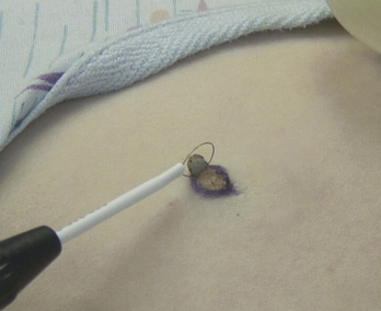
Electrosurgery is a powerful method in performing medically necessary and cosmetic skin procedures in the office setting. The most common cases involve the removal of moles and spider veins, or telangiectasias (Figure 9-1).
Equipment
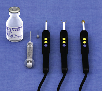
A radiofrequency or electrocautery device provides a wide variety of common clinical uses. In the outpatient setting, mole removal, spider vein ablation, and telangiectasia ablation are useful skills. There are multiple devices on the market with similar uses, although the technology differences may provide different effects when the tools are used. The device has a variety of tips; straight wire, loops, and ball tips are needed for these procedures (Figure 9-2). To assist in identifying the margins of the mole, a surgical marker can be used to highlight the lesion to be removed. This is especially useful for flat nevi, where the margins are not elevated.
Key steps
Cosmetic Mole Removal
Stay updated, free articles. Join our Telegram channel

Full access? Get Clinical Tree


