TECHNIQUE |
FINDINGS |
|---|
EARS |
Inspect auricles and mastoid area |
Examine lateral and medial surfaces and surrounding tissue. |
|
|
|
 Size/shape/symmetry Size/shape/symmetry |
EXPECTED:Familial variations. Auricles of equal size and similar appearance. Darwin tubercle. |
UNEXPECTED:Unequal size or configuration. Cauliflower ear and other deformities. |
 Lesions Lesions |
UNEXPECTED:Moles, cysts or other lesions, nodules, or tophi. |
 Color Color |
EXPECTED:Same color as facial skin. |
UNEXPECTED:Blueness, pallor, or excessive redness. |
 Position Position
Draw imaginary line between inner canthus and most prominent protuberance of occiput. Draw imaginary line perpendicular to first line and anterior to auricle. |
EXPECTED:Top of auricle touching or above horizontal line. Vertical position. |
UNEXPECTED:Auricle positioned below line (low-set); unequal alignment. Lateral posterior angle greater than 10 degrees. |
 Preauricular area Preauricular area |
EXPECTED:Preauricular pits, skin tags, or smooth skin. |
UNEXPECTED:Openings in preauricular area, discharge. |
 External auditory canal External auditory canal |
EXPECTED:No discharge, no odor; canal walls pink. |
UNEXPECTED:Serous, bloody, or purulent discharge; foul smell. |
Palpate auricles and mastoid area |
|
EXPECTED:Firm and mobile, readily recoils from folded position; no tenderness in postauricular or mastoid area. |
UNEXPECTED:Tenderness, swelling, nodules. Pain when pulling on lobule. |
Inspect auditory canal with otoscope |
|
EXPECTED:Minimal cerumen in varying color and texture. Uniformly pink canal. Hairs in outer third of canal. |
UNEXPECTED:Cerumen obscures tympanic membrane, odor, lesions, discharge, scaling, excessive redness, foreign body. |
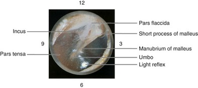
Inspect tympanic membrane |
 Landmarks Landmarks
Vary light direction to observe entire membrane and annulus. |
EXPECTED:Visible landmarks (umbo, handle of malleus, light reflex). |
UNEXPECTED:Perforations, landmarks not visible. |
 Color Color |
EXPECTED:Translucent, pearly gray. |
UNEXPECTED:Amber, yellow, blue, deep red, chalky white, dull, white flecks, or dense white plaques; air bubbles or fluid level. |
|
|
Contour |
EXPECTED:Slightly conical with concavity at umbo. |
UNEXPECTED:Bulging (more conical, usually with loss of bony landmarks and distorted light reflex) or retracted (more concave, usually with |
|
accentuated bony landmarks and distorted light reflex). |
Mobility
Seal canal with speculum, and gently apply positive (squeeze) and negative (release) pressure with pneumatic attachment. |
EXPECTED:Movement in and out. |
UNEXPECTED:No movement. |
Assess hearing |
 Questions during history Questions during history |
EXPECTED:Responds to questions appropriately. |
UNEXPECTED:Excessive requests for repetition. Speech with monotonous tone and erratic volume. |
 Whispered voice Whispered voice
Have patient mask hearing in one ear by inserting a finger in ear canal. Stand 1 to 2 feet from other ear and softly whisper three letter and number combinations (e.g., 3, T, 9 or 5, M, 2). Use a different letter number combination in other ear. |
EXPECTED:Patient repeats numbers and letters correctly more than 50% of time. |
UNEXPECTED:Patient unable to repeat whispered words. |
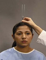
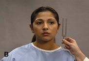
 Weber test Weber test
Place base of vibrating tuning fork on midline vertex of head. Repeat with one ear occluded. |
EXPECTED:Sound heard equally in both ears (unoccluded). Sound heard better in occluded ear. |
UNEXPECTED:See table on p. 84. |
|
|


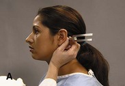
 Rinne test
 Shape/size
 Color
Color Nares
Nares Color
Color Shape
Shape Condition

 Get Clinical Tree app for offline access