Problem 12 Dysphagia and weight loss in a middle-aged man
A 55-year-old man presents with a 3-month history of dysphagia and weight loss. He reports that his initial problem was with solid foods only, but he has had to switch to a minced diet for the past month. His wife uses a blender to prepare all of his meals. He has lost almost 15 kg in weight. He has never been diagnosed with gastro-oesophageal reflux disease, and he does not have any medical problems that he is aware of. He used to smoke a pack of cigarettes per day but quit 5 years ago. He drinks between 6 and 12 beers per week and is not on any medication. In the past he has had an appendicectomy and a right inguinal hernia repair with mesh.
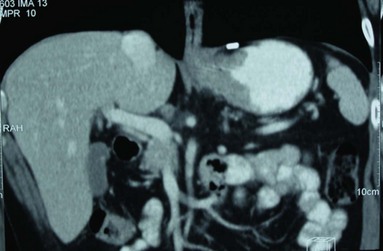
A further investigation is performed and a representative film is shown (Figure 12.1).
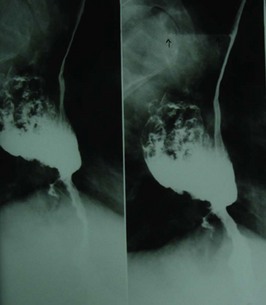
Based on the radiological findings, the patient is referred for another investigation (Figure 12.2).
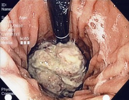
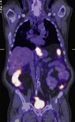
The lesion is biopsied and confirmed to be a moderately differentiated adenocarcinoma. The patient is referred for further staging investigations (Figure 12.3 and Figure 12.4.).
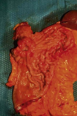
Following three cycles of chemotherapy (over 3 months) and 4 weeks of stabilization, the patient underwent surgery. Figure 12.5 shows the operative view of the upper abdomen.