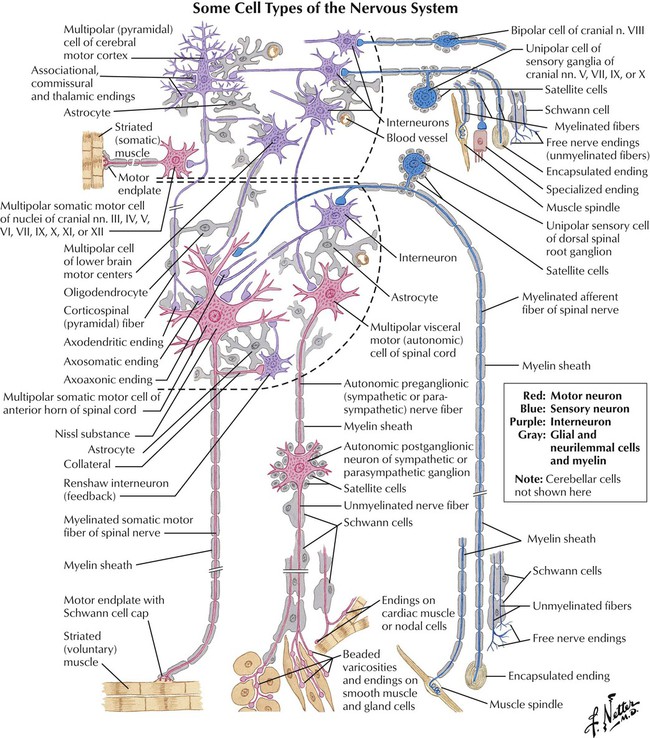
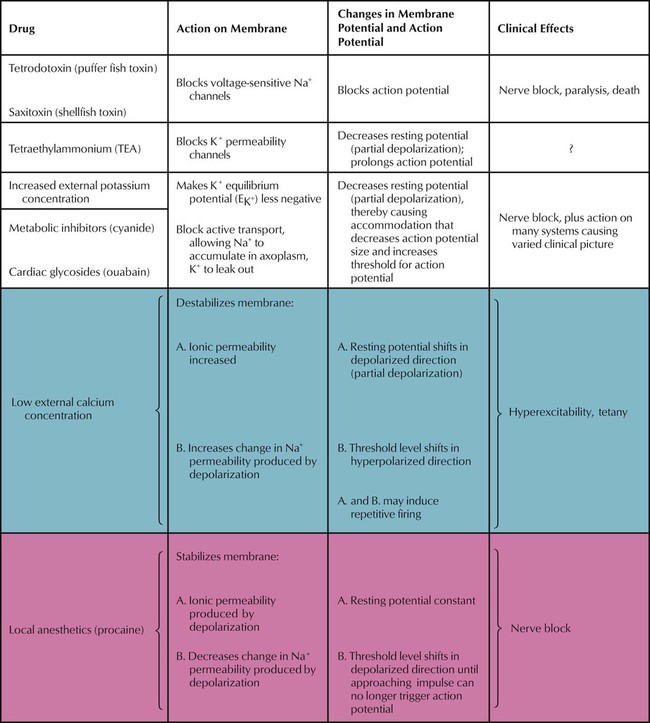
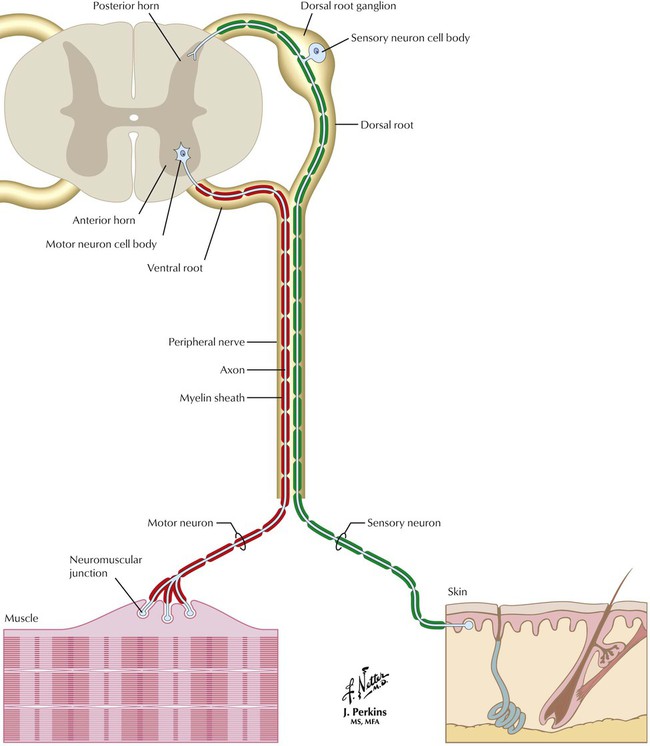
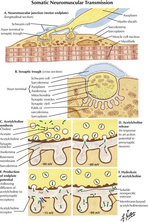
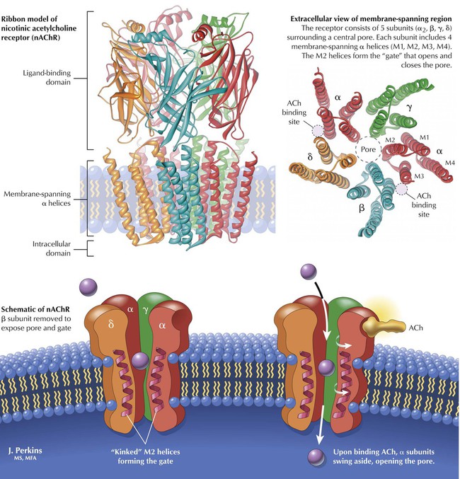
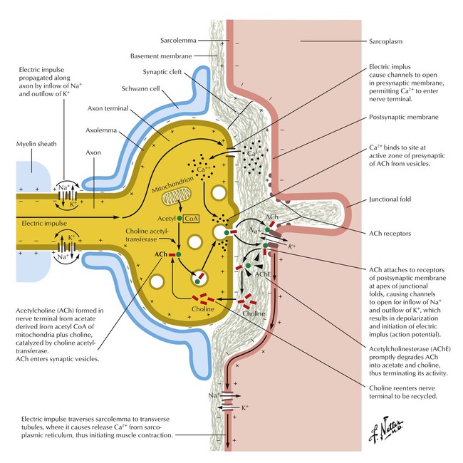
Chapter 2 Drugs Used to Affect the Autonomic and Somatic Nervous Systems Overview The nervous system functions as a major communication system within the body. Information is transmitted by electrical conduction along axons of neurons to (via afferent nerves) and from (via efferent nerves) the central nervous system (CNS). Between neurons or between neurons and target cells are gaps termed synapses across which the signal is transmitted chemically rather than electrically (with some exceptions). The endogenous chemical substances that transmit these signals are termed neurotransmitters. Accuracy of signal transmission requires that the postsynaptic cell reliably receive the intended message from the presynaptic cell. The fidelity is ensured by neurotransmitter-specific receptors located on the postsynaptic cell membrane. Because an action potential, or the change in membrane potential occurring in excitable tissue during excitation, relies on a chemical process (ion flux across the membrane) and the transmission across synapses is primarily chemical, exogenously administered chemicals or drugs can modify physiologic processes mediated by the nervous system. The major neurotransmitters in the periphery are acetylcholine (ACh) and norepinephrine, and drugs can be designed either to mimic or to inhibit their actions. The integrated arrangement of the nervous system and the special distribution of neurotransmitter receptors allow for a targeted drug effect. In most cases, the actual action of the drug—and even much of its unwanted action—is predictable on the basis of the anatomy and physiology of the nervous system. It is convenient for the understanding of drug action to subclassify the peripheral nervous system (PNS) into 2 components: the somatic nervous system (SNS) and the autonomic nervous system (ANS). The nerves of the SNS innervate skeletal muscles, and drugs that act on this system thus affect skeletal muscle function such as tone (eg, muscle relaxants given before surgery). Because all skeletal neuromuscular junctions contain ACh as the neurotransmitter, ACh and its receptors are targets for drugs intended to modify skeletal muscle function. The cholinergic receptors at these skeletal neuromuscular junctions are sufficiently different structurally (3-dimensional shape) from those at other sites to allow drugs to be designed to bind to only this type (nicotinic) of cholinergic receptor. The nerves of the ANS innervate the organs of the body and can be further classified into sympathetic and parasympathetic subdivisions. Sympathetic activity is increased by drugs that mimic or enhance the action of norepinephrine. Parasympathetic activity is increased by drugs that mimic or enhance the action of ACh. Both systems are tonically active. Hence, antagonism of one system results in enhanced activity of the other. The SNS and ANS together provide a mechanistic framework for understanding the effects (good and bad) of drugs. Elucidation of additional roles for neurotransmitters and identification of other receptor subtypes will likely lead to development of more selective drugs. Such drugs will be found by using, for example, high-throughput screening assays or molecular modeling techniques—or even by serendipity. However they are discovered, they should permit more selective targeting of the therapeutic end point with fewer unwanted effects. Figure 2-1 Organization of the Nervous SystemThe actions of many drugs can be understood as the modulation of the nervous system’s control of physiologic processes. The CNS and PNS communicate via afferent and efferent neurons. As a result of this anatomical organization, drugs can affect sensory input (eg, local anesthetics for pain), skeletal muscle activity (eg, muscle relaxants for surgery), or autonomic output (eg, drugs that act on blood vessels or the heart to reduce high blood pressure). Figure 2-2 Action of Drugs on Nerve ExcitabilityEfficient and effective transmission of neuronal action potentials relies on the unequal distribution of positive (primarily Na+ and K+) and negative (primarily Cl−) ions across the axonal membrane. Selective, voltage-sensitive permeability of the membrane to these ions establishes the unequal distribution of the ions according to the Nernst equation and gives rise to a resting transmembrane potential difference. Drugs that alter the ion flux affect the resting transmembrane potential difference. The larger this difference, the further the neuron is from its firing threshold and the less likely that it will fire (ie, initiate an action potential). The smaller the transmembrane potential difference, the more likely it is that the neuron will reach this threshold and fire. Figure 2-3 Interface of the Central and Peripheral Nervous Systems and Organization of the Somatic DivisionSpinal nerve pairs enter and exit along segmented caudal, thoracic, lumbar, and sacral portions of the spinal cord and distribute throughout the body. Somatic afferent neurons transmit sensory information about normal status (eg, proprioception) or pathologic states (eg, heat and mechanical damage) to the spinal cord and brain. Efferent neurons carry motor signals from the spinal cord and brain to the somatic (striated or skeletal muscles: effectors) and autonomic (smooth muscle, cardiac muscle, glands) divisions of the PNS. Drugs can selectively modulate the activity of afferent or efferent pathways: those that excite afferent nociceptive neurons produce pain; those that inhibit afferent nociceptive neurons are analgesic. Those that excite efferent, or neuromuscular, junctions produce tetanus; those that inhibit these junctions cause paralysis. Figure 2-4 Neuromuscular TransmissionNeurons innervate skeletal muscles at the neuromuscular junction (A). The axon-muscle interface forms at a synaptic trough, which has extensive foldings that increase the surface area of exposure to a neurotransmitter (B). ACh, the neurotransmitter at neuromuscular junctions, is synthesized in the presynaptic neuron from mitochondrial acetyl-CoA and extracellular choline via an enzyme-catalyzed reaction. ACh is stored in presynaptic vesicles (C) until release in response to an action potential in the presynaptic neuron (D), a Ca2+-dependent process. ACh diffuses across the synaptic cleft and binds reversibly to specific receptor sites on the postsynaptic membrane. Ion flux then increases and the postsynaptic membrane depolarizes (E), which triggers an action potential that leads to muscle contraction. Released ACh is eliminated from the synapse by cholinesterase action (F). Figure 2-5 Nicotinic Acetylcholine ReceptorDrugs that block cholinesterases prolong the ACh residency time in the synapse and enhance the effect of ACh. Receptors at neuromuscular junctions are termed nicotinic cholinergic receptors (nAChRs) because nicotine is a relatively selective agonist at these sites. In an nAChR, 5 subunits (α2, β, γ, σ) form a cluster around a central cation-selective pore. Two ACh-binding sites are in the extracellular part of the receptor between α and the other subunits. When ACh binds to the sites, the receptor conformation changes: α subunits swing out, and the channel opens. Charged amino acids lining the pore select ions that can pass into the cell. Figure 2-6 Physiology of the Neuromuscular JunctionAs Loewi demonstrated in the 1920s, a gap (synapse) exists between an ANS neuron’s axon terminal and the adjacent neuron or effector cell. Information is transmitted across this gap via chemical transmitters (neurotransmission). Neurotransmitters are commonly stored in presynaptic vesicles; arrival of an action potential stimulates a Ca2+-dependent neurotransmitter release into the synapse. The neurotransmitter crosses the gap and binds to highly selective receptor molecules on the postsynaptic cell, thereby modifying the activity of the postsynaptic cell. Neurotransmission provides fidelity of signal transmission. ANS neurotransmitters are simple organic molecules, and exogenous chemicals (drugs) can modify (mimic or antagonize) the action of the endogenous ANS neurotransmitters. < div class='tao-gold-member'> Only gold members can continue reading. Log In or Register a > to continue Share this:Click to share on Twitter (Opens in new window)Click to share on Facebook (Opens in new window)Like this:Like Loading... Related Related posts: Drugs Used in Neoplastic Disorders Drugs Used in Disorders of the Central Nervous System and Treatment of Pain Drugs Used in Infectious Disease Drugs Used in Disorders of the Respiratory System Stay updated, free articles. Join our Telegram channel Join Tags: Netters Illustrated Pharmacology Updated Edition with Student Jun 21, 2016 | Posted by admin in PHARMACY | Comments Off on Drugs Used to Affect the Autonomic and Somatic Nervous Systems Full access? Get Clinical Tree
Chapter 2 Drugs Used to Affect the Autonomic and Somatic Nervous Systems Overview The nervous system functions as a major communication system within the body. Information is transmitted by electrical conduction along axons of neurons to (via afferent nerves) and from (via efferent nerves) the central nervous system (CNS). Between neurons or between neurons and target cells are gaps termed synapses across which the signal is transmitted chemically rather than electrically (with some exceptions). The endogenous chemical substances that transmit these signals are termed neurotransmitters. Accuracy of signal transmission requires that the postsynaptic cell reliably receive the intended message from the presynaptic cell. The fidelity is ensured by neurotransmitter-specific receptors located on the postsynaptic cell membrane. Because an action potential, or the change in membrane potential occurring in excitable tissue during excitation, relies on a chemical process (ion flux across the membrane) and the transmission across synapses is primarily chemical, exogenously administered chemicals or drugs can modify physiologic processes mediated by the nervous system. The major neurotransmitters in the periphery are acetylcholine (ACh) and norepinephrine, and drugs can be designed either to mimic or to inhibit their actions. The integrated arrangement of the nervous system and the special distribution of neurotransmitter receptors allow for a targeted drug effect. In most cases, the actual action of the drug—and even much of its unwanted action—is predictable on the basis of the anatomy and physiology of the nervous system. It is convenient for the understanding of drug action to subclassify the peripheral nervous system (PNS) into 2 components: the somatic nervous system (SNS) and the autonomic nervous system (ANS). The nerves of the SNS innervate skeletal muscles, and drugs that act on this system thus affect skeletal muscle function such as tone (eg, muscle relaxants given before surgery). Because all skeletal neuromuscular junctions contain ACh as the neurotransmitter, ACh and its receptors are targets for drugs intended to modify skeletal muscle function. The cholinergic receptors at these skeletal neuromuscular junctions are sufficiently different structurally (3-dimensional shape) from those at other sites to allow drugs to be designed to bind to only this type (nicotinic) of cholinergic receptor. The nerves of the ANS innervate the organs of the body and can be further classified into sympathetic and parasympathetic subdivisions. Sympathetic activity is increased by drugs that mimic or enhance the action of norepinephrine. Parasympathetic activity is increased by drugs that mimic or enhance the action of ACh. Both systems are tonically active. Hence, antagonism of one system results in enhanced activity of the other. The SNS and ANS together provide a mechanistic framework for understanding the effects (good and bad) of drugs. Elucidation of additional roles for neurotransmitters and identification of other receptor subtypes will likely lead to development of more selective drugs. Such drugs will be found by using, for example, high-throughput screening assays or molecular modeling techniques—or even by serendipity. However they are discovered, they should permit more selective targeting of the therapeutic end point with fewer unwanted effects. Figure 2-1 Organization of the Nervous SystemThe actions of many drugs can be understood as the modulation of the nervous system’s control of physiologic processes. The CNS and PNS communicate via afferent and efferent neurons. As a result of this anatomical organization, drugs can affect sensory input (eg, local anesthetics for pain), skeletal muscle activity (eg, muscle relaxants for surgery), or autonomic output (eg, drugs that act on blood vessels or the heart to reduce high blood pressure). Figure 2-2 Action of Drugs on Nerve ExcitabilityEfficient and effective transmission of neuronal action potentials relies on the unequal distribution of positive (primarily Na+ and K+) and negative (primarily Cl−) ions across the axonal membrane. Selective, voltage-sensitive permeability of the membrane to these ions establishes the unequal distribution of the ions according to the Nernst equation and gives rise to a resting transmembrane potential difference. Drugs that alter the ion flux affect the resting transmembrane potential difference. The larger this difference, the further the neuron is from its firing threshold and the less likely that it will fire (ie, initiate an action potential). The smaller the transmembrane potential difference, the more likely it is that the neuron will reach this threshold and fire. Figure 2-3 Interface of the Central and Peripheral Nervous Systems and Organization of the Somatic DivisionSpinal nerve pairs enter and exit along segmented caudal, thoracic, lumbar, and sacral portions of the spinal cord and distribute throughout the body. Somatic afferent neurons transmit sensory information about normal status (eg, proprioception) or pathologic states (eg, heat and mechanical damage) to the spinal cord and brain. Efferent neurons carry motor signals from the spinal cord and brain to the somatic (striated or skeletal muscles: effectors) and autonomic (smooth muscle, cardiac muscle, glands) divisions of the PNS. Drugs can selectively modulate the activity of afferent or efferent pathways: those that excite afferent nociceptive neurons produce pain; those that inhibit afferent nociceptive neurons are analgesic. Those that excite efferent, or neuromuscular, junctions produce tetanus; those that inhibit these junctions cause paralysis. Figure 2-4 Neuromuscular TransmissionNeurons innervate skeletal muscles at the neuromuscular junction (A). The axon-muscle interface forms at a synaptic trough, which has extensive foldings that increase the surface area of exposure to a neurotransmitter (B). ACh, the neurotransmitter at neuromuscular junctions, is synthesized in the presynaptic neuron from mitochondrial acetyl-CoA and extracellular choline via an enzyme-catalyzed reaction. ACh is stored in presynaptic vesicles (C) until release in response to an action potential in the presynaptic neuron (D), a Ca2+-dependent process. ACh diffuses across the synaptic cleft and binds reversibly to specific receptor sites on the postsynaptic membrane. Ion flux then increases and the postsynaptic membrane depolarizes (E), which triggers an action potential that leads to muscle contraction. Released ACh is eliminated from the synapse by cholinesterase action (F). Figure 2-5 Nicotinic Acetylcholine ReceptorDrugs that block cholinesterases prolong the ACh residency time in the synapse and enhance the effect of ACh. Receptors at neuromuscular junctions are termed nicotinic cholinergic receptors (nAChRs) because nicotine is a relatively selective agonist at these sites. In an nAChR, 5 subunits (α2, β, γ, σ) form a cluster around a central cation-selective pore. Two ACh-binding sites are in the extracellular part of the receptor between α and the other subunits. When ACh binds to the sites, the receptor conformation changes: α subunits swing out, and the channel opens. Charged amino acids lining the pore select ions that can pass into the cell. Figure 2-6 Physiology of the Neuromuscular JunctionAs Loewi demonstrated in the 1920s, a gap (synapse) exists between an ANS neuron’s axon terminal and the adjacent neuron or effector cell. Information is transmitted across this gap via chemical transmitters (neurotransmission). Neurotransmitters are commonly stored in presynaptic vesicles; arrival of an action potential stimulates a Ca2+-dependent neurotransmitter release into the synapse. The neurotransmitter crosses the gap and binds to highly selective receptor molecules on the postsynaptic cell, thereby modifying the activity of the postsynaptic cell. Neurotransmission provides fidelity of signal transmission. ANS neurotransmitters are simple organic molecules, and exogenous chemicals (drugs) can modify (mimic or antagonize) the action of the endogenous ANS neurotransmitters. < div class='tao-gold-member'> Only gold members can continue reading. Log In or Register a > to continue Share this:Click to share on Twitter (Opens in new window)Click to share on Facebook (Opens in new window)Like this:Like Loading... Related Related posts: Drugs Used in Neoplastic Disorders Drugs Used in Disorders of the Central Nervous System and Treatment of Pain Drugs Used in Infectious Disease Drugs Used in Disorders of the Respiratory System Stay updated, free articles. Join our Telegram channel Join