Fig. 17.1
Structure of neuromuscular junction (With permission from Oxford University Press; Br J Anaesth CEPD Rev. 2002;2(5):129–33)
The area of the nerve which lies closest to the muscle cell is known as the synapse. The motor end plate is a specialized area in the muscle cell which fronts the nerve. The synapse and the end plate are separated by a gap (approximately 20 nm) called the synaptic or junctional cleft. The gap is filled with extracellular fluid. As the action potential arrives at the synapse, it causes the neurotransmitter acetylcholine to be released. Acetylcholine then travels across the synaptic cleft and binds selectively to the acetylcholine receptor at the post-synaptic motor end plate of the muscle, causing an action potential to travel through it.
The amount of acetylcholine released following a nerve action potential is far in excess of what is needed to reach the threshold at the end plate. The acetylcholine receptor acts like a switch where it remains closed until the acetylcholine binds to it. It then opens to allow current to pass through it. It closes immediately when the acetylcholine detaches from the receptor.
Acetylcholinesterase
Acetylcholine molecules that do not react with a receptor or are released from the binding site are destroyed almost immediately by the enzyme acetylcholinesterase in the junctional cleft. There are two distinct types of cholinesterase, namely acetylcholinesterase (AChE) and butyrylcholinesterase (BChE). They are closely related in molecular structure but differ in their distribution, substrate specificity and functions. AChE is mainly membrane bound, relatively specific for acetylcholine and responsible for its rapid hydrolysis at cholinergic synapses. BChE or pseudocholinesterase is relatively non-selective and is found in many tissues. It hydrolyses butyrylcholine more rapidly than acetylcholine as well as other esters such as procaine and succinylcholine.
Conditions That Manifest as Acute Neuromuscular Weakness
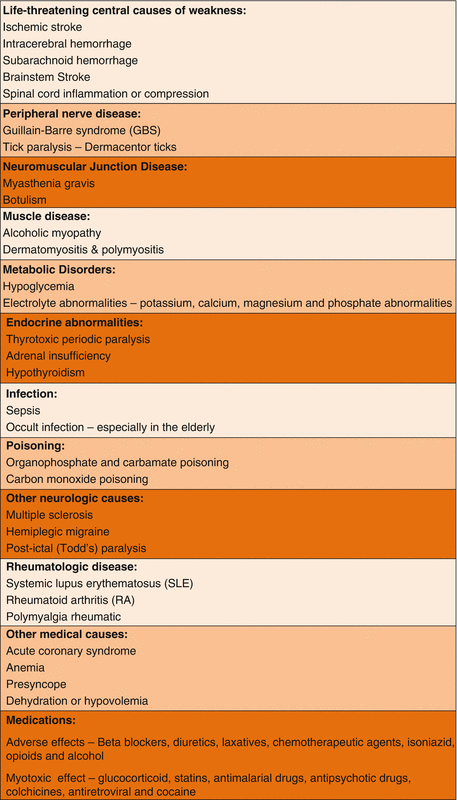
There are many conditions that can cause acute muscular weakness or paralysis. In primary neuromuscular disorders, the pathology can occur centrally in the brain down to the peripheral nerves including the neuromuscular junctions, and the muscle itself. Many systemic disorders, as shown in Table 17.1, can also bring about muscle weakness.

Table 17.1
Conditions causing muscle weakness or paralysis

Drugs Used to Treat Primary Neuromuscular Diseases
Drugs used to treat primary neuromuscular diseases work at different sites, depending on the pathology. In myasthenia gravis there is a transmission failure in the neuromuscular junction. This is due to an autoimmune response that destroys the nicotinic acetylcholine receptors. Drugs that can enhance cholinergic transmission, such as anticholinesterases, can improve neuromuscular function in this condition. However if the disease is too severe, the number of receptors remaining may become too few to produce an adequate end-plate potential, thus rendering anticholinesterase treatment ineffective. Other treatment modalities for myasthenia gravis involve the use of corticosteroids, intravenous immunoglobulins and plasmapheresis, with the aim of suppressing the amount of circulating immunoglobulins which attack the ACh receptors.
Drugs That Enhance Cholinergic Transmission
Cholinergic transmission can be enhanced either by inhibiting cholinesterase or by increasing acetylcholine release. The peripherally acting anticholinesterase drugs are mainly used for neuromuscular disorders, whereas the centrally acting anticholinesterase agents have been found to be effective in the treatment of neuropsychiatric disorders such as dementia. There are no clinically useful drugs which increase the release of ACh.
Short-acting Anticholinesterase
These agents are used to diagnose myasthenia gravis (Tensilon test) or to determine the effectiveness of treatment with anticholinesterase therapy
Stay updated, free articles. Join our Telegram channel

Full access? Get Clinical Tree


