Fig. 11.1
The mechanism by which catecholamine exerts its effects. Gs-GTP: Stimulating Guanosine nucleotide-binding protein-Guanosine Triphosphate complex; cAMP: cyclic Adenosine Monophosphate; Ca2+: Calcium; SR: sarcoplasmic reticulum (Reproduced with permission from Wolters Kluwer Health; Circulation 2008; 118: 1047–56)
Dopamine produces different effects at different doses. At low doses it acts mainly on dopaminergic receptors, giving rise to positive inotropy. β-adrenergic effects (positive inotropy and chronotropy) followed by α effects (increased systemic vascular resistance (SVR)) are seen as the dose increases, with predominantly α effects at high doses causing marked increase in SVR.
Dobutamine is a synthetic sympathomimetic. It is also administered via intravenous infusion. It mainly affects β-adrenoceptors, causing positive inotropy, and has variable effects on vascular resistance and heart rate.
Adrenaline has both α and β adrenergic effects, causing positive inotropy, chronotropy and increased SVR.
2.
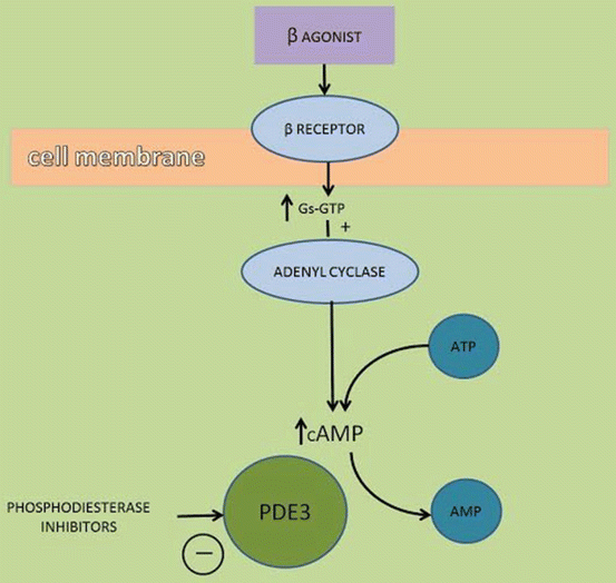
Phosphodiesterase-III inhibitors (PDIs): these drugs work inside the cell to prevent the breakdown of cAMP, thereby increasing intracellular Ca2+ concentrations (Fig. 11.2). A commonly used drug in this category is milrinone.


Fig. 11.2
Mechanism by which Phosphodiesterase inhibitors work to increase intracellular calcium concentrations. Gs-GTP: Stimulating Guanosine nucleotide-binding protein-Guanosine triphosphate complex; cAMP: cyclic adenosine monophosphate; ATP: Adenosine triphosphate; PDE3: Phosphodiesterase 3 (Reproduced with permission from Wolters Kluwer Health; Circulation 2008; 118: 1047–56)
Milrinone is also administered via intravenous infusion. It is also called an inodilator, as it has positive inotropic effects while giving rise to reduced SVR. It is metabolised in the liver.
3.
Digitalis: This glycoside inhibits Na+/K+-ATPase thus increasing the intracellular sodium concentrations. This in turn increases intracellular calcium concentrations (via the Na+/Ca2+ exchanger), augmenting the actin-myosin interaction and giving rise to stronger contractions. This also increases the action potential (AP) duration and the refractory period of the atrioventricular (AV) node and the bundle of His, thereby reducing AV conductivity and reducing ventricular rate, making it useful in the treatment of arrhythmias. A common digitalis is digoxin.
Coronary Vasodilators
Pump dysfunction may be due to myocardial ischemia caused by oxygen supply–demand imbalance. Caregivers attempt to maintain blood supply to the myocardium and reduce its oxygen requirement to ensure adequate oxygen supply. This in turn will improve its pump function. One method used to maintain the myocardial oxygen demand and supply balance is by administering coronary vasodilators, i.e., nitrates.
Glyceryl trinitrate (GTN) is a direct acting vasodilator that is metabolised to nitric oxide (nitric oxide donor). Activation of M3 receptors in blood vessels increases nitric oxide synthesis which then diffuses into smooth muscle cells and increases cyclic GMP. This acts on myosin light chain phosphatase that causes inactivation of myosin light chain. The resulting smooth muscle relaxation causes vasodilation. Dilatation of epicardial and interconnecting coronary arteries increases coronary blood flow both in normal and stenotic vessels. At low doses, the effect of GTN is seen in capacitance vessels (veins), affecting preload (venous return) more than afterload (SVR). This reduces left ventricular end diastolic pressures (LVEDP) and myocardial wall tension, increasing sub-endocardial blood flow. Care must be taken, however, as at high doses GTN causes a reduction in the SVR as well. This will in turn reduce coronary perfusion pressure (aortic pressure).
GTN is indicated in the treatment of myocardial ischemia (during acute coronary syndrome), hypertensive crises, heart failure and coronary vasospasm. It is only suitable for titrated intravenous infusions (0.1–10 mcg/kg/min), sublingual delivery (300–500 mcg) and transdermal application (5–10 mg/24 h). It has immediate onset and offset. t½β is 1–4 min. It is mostly metabolised in the liver to both active and inactive metabolites. Adverse reactions include tolerance, rebound hypertension, increased intrapulmonary shunt, loss of hypoxic pulmonary vasoconstriction and methemoglobinemia. Symptoms include tachycardia, dizziness, drowsiness, vertigo, facial flushing, weakness and fainting.
Antiarrhythmia Drugs
The optimal rhythm for maximal efficiency of pump function is sinus rhythm. In this state, there is synchronized and timely flow of blood from the atrium to the ventricles, which is lost in the presence of arrhythmias. Caregivers attempt to control cardiac rhythm, failing which they try to control the rate so that there will still be adequate filling to ensure reasonable output, e.g., in atrial fibrillation, rate control is often used.
Drugs that maintain sinus rhythm or control the heart rate are called anti-arrhythmia agents. A popular classification of these drugs is the Vaughn Williams classification. Class I and IV acts on ion channels, Class II on receptors and Class III acts on both. Most of these drugs have mechanisms of action which overlap.
Class I Antiarrhythmic Drugs
These are sodium channel blockers. They are further divided into three groups.
Class Ia prolongs action potential duration and reduces AV conduction rate. An example is procainamide, used in the treatment of ventricular arrhythmia and atrial fibrillation (AF), administered intravenously or orally. It is metabolized by the liver, with an active metabolite (NAPA, a potassium channel blocker). Its t1/2 is 3–5 h. Common side effects include ventricular arrhythmias, including torsades de pointes, lupus like syndromes and blood dyscrasias.
Class Ib agents reduce action potential duration, but have no effect on AV conduction rate. An example is lignocaine, which is indicated in ventricular arrhythmias. Its route of administration is intravenous and its t1/2 is 1.5–2 h. Common side effects include tremors, paraesthesia and seizures.
Class Ic agents reduce AV conduction rate and have no effect on action potential duration. An example is flecainide, which is indicated in the treatment of paroxysmal atrial fibrillation and Wolf-Parkinson-White syndrome. Its route of administration is oral. It undergoes hepatic metabolism and its t1/2 is 20 h. It is contraindicated in patients with structural heart disease, and side effects include arrhythmias (especially in post-myocardial infarct patients), heart failure, headache and blurred vision.
Class II Antiarrhythmic Drugs
These are β-receptor antagonists. They bind to β1-adrenoceptors located in cardiac nodal tissue, the conducting system, and contracting myocytes, inhibiting the effects of adrenaline on the β1-adrenoceptors. Blockade of β-adrenoceptors inhibits activation of calcium channels, leading to a reduction of AV node conduction. One example is esmolol, which is used in the treatment of paroxysmal supraventricular tachycardia, rate control in non-pre-excited AF or atrial flutter, ectopic atrial tachycardia, inappropriate sinus tachycardia, polymorphic ventricular tachycardia (VT) due to torsades de pointes or myocardial ischemia. It is administered intravenously.
Stay updated, free articles. Join our Telegram channel

Full access? Get Clinical Tree


