Chemical and Cellular Physiology
An atom is the most basic structure of matter. It is composed of a nucleus that contains positively charged protons and uncharged neutrons and negatively charged electrons that revolve around the nucleus much like the planets travel around the sun.
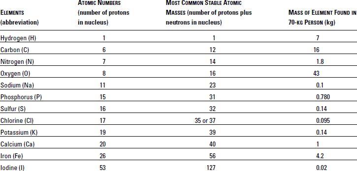
Atoms are defined by the number of protons in their nuclei. As shown in Figure 4-1, carbon has six protons. (One proton is not visible in the figure.) If that atom has seven protons, it would no longer be carbon. Instead, it would be nitrogen (Table 4-1). The number of protons in the nucleus is also referred to as the atomic number. An element is made up of only one type of atom and cannot be further broken down by ordinary chemical means. Ninety-six percent of the body is composed of just four elements—oxygen, carbon, hydrogen, and nitrogen.
In addition to an atomic number, each element has an atomic mass number, which is the sum of its protons and neutrons. For example, carbon (abbreviated as “C”) has a mass number of 12, meaning that each atom in the element has a total of 12 neutrons and protons in its nucleus.
While elements generally have an equal number of protons and neutrons, not all do. In fact, the presence of one or more extra neutrons can result in an unstable configuration. These unstable atoms break down by sending out, or emitting, radioactive particles that over time transform the atom to the more stable configuration. Chapter 10 includes more information on medications that contain radioactive elements.
A molecule is produced when two or more atoms share electrons or combine with each other through a type of magnetic attraction. For example, a molecule of water is designated H2O, meaning that two atoms of hydrogen (H) share electrons with one atom of oxygen (O).
Biological systems are made up largely of molecules that can be classified as sugars (carbohydrates), fats (lipids), and proteins (which often help make chemical reactions occur and are therefore called enzymes). Carbohydrates, fats, and proteins make up most of the human diet, along with vitamins and minerals such as calcium and iron.
Amino acids—20 of them—are the building blocks of proteins. Proteins can be relatively simple molecules, such as insulin, or can be very complex structures, and their effects on cells are often striking.
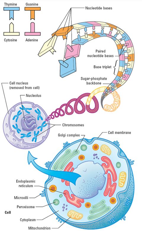
A cell is a living structural and functional unit surrounded by a membrane that has many substructures, as illustrated in Figure 4-2. At the center of each cell is the nucleus, which contains genetic material. As cells divide, each cell contains the same genetic material. Chromosomes are long strands of DNA, an abbreviation for deoxyribonucleic acid. In DNA, the sugar deoxyribose forms a kind of backbone in combination with phospate (phosphorus plus oxygen), and this backbone supports paired nucleotide bases composed of either adenine–thymine or cytosine–guanine. Each person has 46 chromosomes in each cell, 23 contributed from each parent.
Sections of these 46 chromosomes that code for proteins are called genes. Genes are characterized by the specific order and pairing of four nucleotide bases. Genes essentially code for the production of proteins within the cell, but some genes also turn on and off the activity of other genes.
| Figure 4-2 | Structure of the Human Cell (bottom), Nucleus of that Cell (middle), and the Genetic Material of the Cell, DNA (top) |
Anatomy and Organ Systems
As you may remember from biology or health courses in high school, the human body is made up of many highly specialized tissues, including bones, muscles, and nerves. Specialized cells are grouped together in organs of the body, such as the lungs or stomach. Organs that interrelate to one another in function are termed organ systems. The body has 11 organ systems, as outlined below.1–2
Many diseases result from disruptions in one or more of these organ systems. Drugs and drug products that are primarily used to treat these diseases are often grouped together, and you should be able to name several agents useful in treating the most common diseases affecting people. The organ system approach is generally used for making these drug categorizations, as shown at the end of this chapter in Table 4-3.
Integumentary System
The integumentary system literally holds the body together and helps it stay cool and interact with its environment. As shown in Figure 4-3, this system includes the skin, hair, nails, and sweat glands and is responsible for protection, body temperature regulation, conservation of water, and production of vitamin D precursors.
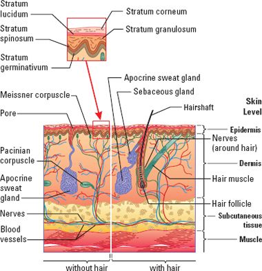
The skin is composed of a thin, outermost area called the epidermis, under which lies the thicker dermis. Just beneath the skin is the subcutaneous tissue, which contains fat cells, blood vessels, and nerves to supply the skin. Sebaceous glands are usually connected to hair follicles and secrete an oily substance called sebum which keeps the skin and hair from drying out. Sweat glands produce around 600 mL of sweat per day, which serves to cool the body as it evaporates. Vitamin D, needed for the absorption of calcium, is made when sunlight activates a precursor located in the skin. The skin is the site of administration for many drugs—those injected into the subcutaneous tissue and those administered by transdermal patch. In both cases, the drug generally reaches the blood and is carried throughout the body.
Skeletal System
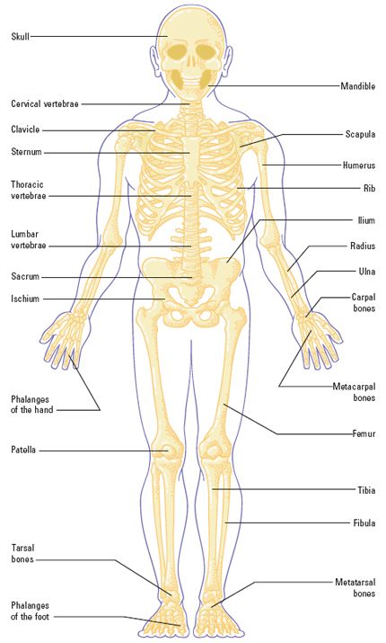
As shown in Figure 4-4, the skeletal system consists of bones, cartilage, and joints. Without the skeletal system, people would be like jellyfish—incapable of initiating movement or picking up items.
Adults have 206 bones, including 106 just in the hands and feet! Bones protect internal organs, allow body movement, and provide attachment for skeletal muscle.
Bones are richly supplied with blood vessels and nerves. The bone marrow, located in the ends of long bones in the arms and legs and in the pelvis, ribs, sternum (breastbone), and skull, produces blood cells. Bone also stores calcium, phosphorus, and triglycerides.
Joints—formed when two bones meet—allow movement. They contain synovial fluid that lubricates the joint.
Cartilage is similar in function to bone but very different in many other ways. Cartilage, which you can feel in your ears and nose, consists of collagen fibers and does not contain blood vessels or nerves. It provides support but allows flexibility.
Muscular System
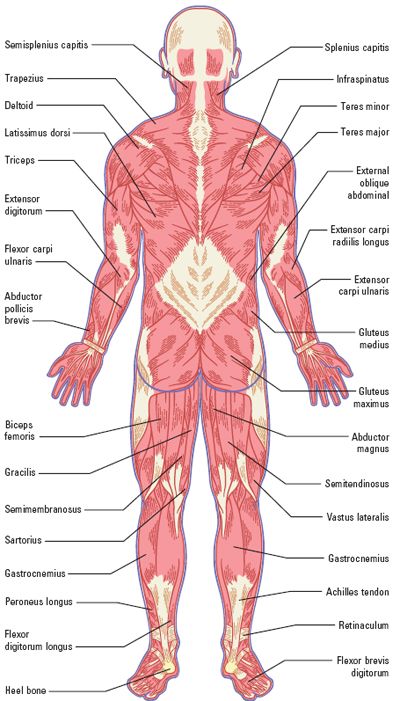
Muscles attach to bones and coordinate body movements, maintain posture, produce body heat, and control outflow of certain liquids (Figure 4-5). Without the muscles and associated tissues (ligaments and tendons), the body would collapse like a floppy puppet.
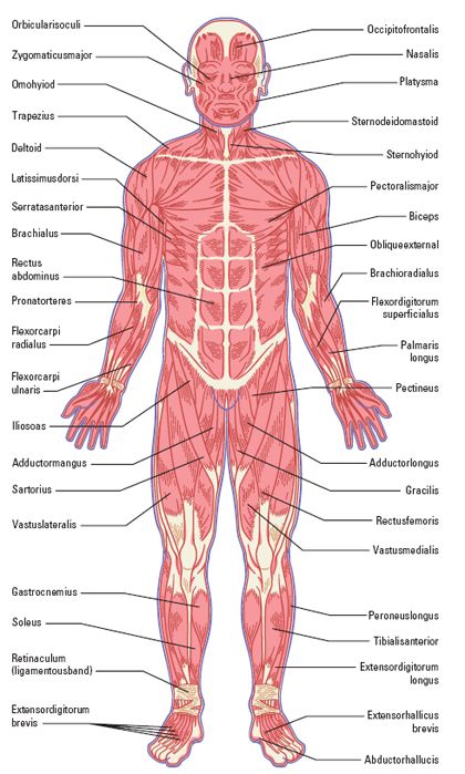
Three types of muscles are found in the human body:
- Skeletal muscle—connects to and moves bones. Impulses from nerves allow voluntary movement. Since the nerve and muscle don’t directly touch, a chemical called a neurotransmitter (acetylcholine) is released from a nerve ending at the junction of the nerve and muscle to direct muscle movement.
- Cardiac muscle—contained in the walls of the heart and causes the heart to contract (beat). Unlike skeletal muscle, contraction is not dependent on neurotransmitters and is autoregulated.
- Smooth muscle—found in blood vessels, airways, the iris of the eye, and in other hollow organs such as the bladder. Like cardiac muscle, smooth muscle is not voluntarily controlled.
Nervous System
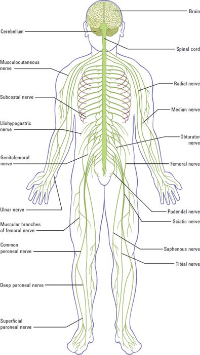
The nervous system, depicted in Figure 4-6, is responsible for gathering information from in and around the body (whether it be a pin prick on the skin or a noise heard or the acidity of the blood), processing this information in the brain and determining how to react, and lastly, conveying the brain’s “decision” back to the muscles, glands, and organs responsible for performing the function. In addition, nerves go to the internal organs of the body to monitor and, in some cases, direct activity.
Nerves that send information to the brain are called afferent nerves, and those that send information from the brain to the tissue are called efferent nerves. The nervous system is composed of the central nervous system (CNS), the brain (including the cerebellum shown in Figure 4-6) and spinal cord, and the peripheral nervous system (PNS)—all of the nerves outside of the CNS. The PNS can be further subdivided as follows:
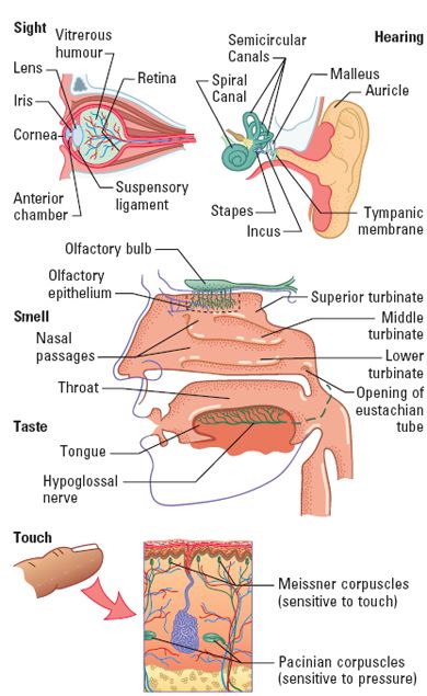
- Somatic nervous system—afferent nerves gather information from the five senses (sight, smell, touch, taste, and sound) and efferent nerves return information to skeletal muscles (Figure 4-7). This system is under voluntary control.
- Autonomic nervous system—afferent nerves gather information from various organs such as the stomach and lungs. Efferent nerves are classified as sympathetic and parasympathetic and generally have opposing actions, like increasing versus decreasing the heart rate. This system is generally involuntary. That is, it operates without the person being aware of or in control of its actions.
Endocrine System
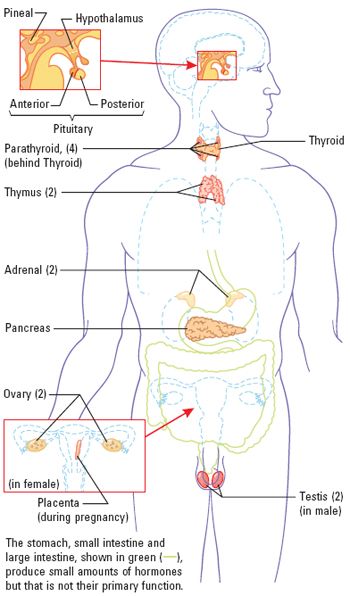
This system includes glands that secrete hormones and related substances that control functions such as metabolism, reproduction, and growth. Hormones are excreted in response to signals originating in the “master” glands—the hypothalamus, a part of the brain, and pituitary glands, located at the base of the brain and connected to the hypothalamus (Figure 4-8).
For example, if blood glucose (sugar) levels are low, the hypothalamus will excrete a hormone to tell the pituitary to secrete a second hormone that tells the pancreas to secrete glucagon and the liver to produce glucose. If blood glucose becomes too high, the hypothalamus secretes an inhibiting hormone that ultimately stops the production of glucose and causes the release of insulin from the pancreas. Thus, hormones are excreted via a chemical feedback mechanism.
The pineal gland in the brain secretes melatonin, which induces sleep. Melatonin is sold in pharmacies and other retail outlets as a dietary supplement that helps people get to sleep.
As shown in Figure 4-8, other glands in the human body include the following:
- Thyroid gland—produces thyroid hormone, which controls cellular metabolism and regulates body temperature.
- Parathyroid gland—regulates body levels of calcium, phosphate, and magnesium.
- Thymus gland—produces hormones that promote immunity (the body’s defense system against infection).
- Adrenal glands—produce glucocorticoids (cortisol), mineralocorticoids (aldosterone), small amounts of androgens, and epinephrine and norepinephrine. Glucocorticoids regulate metabolism and resistance to stress and have anti-inflammatory effects. Mineralocorticoids regulate blood pressure by affecting the amount of fluid retained in the circulation. Epinephrine and norepinephrine, also known as adrenaline and noradrenaline, respectively, are excreted during exercise or in stressful situations to increase heart rate and blood flow to various organs and to dilate the airways.
- Pancreas—secretes insulin, which lowers blood sugar (glucose) levels, and glucagon, which raises blood glucose levels.
- Ovaries (in girls and women)—produce estrogen and progesterone, which regulate the menstrual cycle, maintain pregnancy and lactation, and promote development of female sexual characteristics.
- Testes (in boys and men)—produce testosterone, needed for sperm production and male sexual characteristics.
Includes the central nervous system (the brain, cerebellum, and spinal cord) and the peripheral nervous system (everything else).
Cardiovascular System
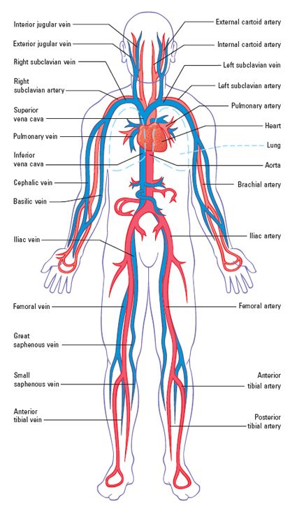
The cardiovascular system consists of the heart, blood vessels, and blood (Figure 4-9). This system is responsible for transporting nutrients, waste products, gases, and hormones; some immune functions; and regulation of temperature. This system has many parts, including the following:
- Heart—The heart contains four chambers and four valves. The right atrium receives blood from the body through the superior and inferior vena cava. It is then pumped through the tricuspid valve into the right ventricle, which pumps blood to the lungs through the pulmonary valve into the right and left pulmonary arteries. From the lungs, oxygenated blood is received back into the heart through the pulmonary veins, into the left atrium. Blood passes through the bicuspid valve into the left ventricle. The left ventricle pumps blood throughout the body through the aortic valve and into the aorta.
- Blood vessels—Blood vessels that carry blood from the heart are called arteries. Arterial blood contains oxygen (except for the blood in the pulmonary arteries, which carry oxygen from the right side of the heart to the lungs where oxygenation occurs). Capillaries are very small blood vessels that connect arteries and veins, and this is where nutrients and gases are exchanged within the tissues. Veins carry blood back to the heart. Since the oxygen has been released into the tissues, venous blood is deoxygenated (except for blood within the pulmonary veins, which go from the lungs to the left side of the heart for pumping to the tissues). The heart muscle itself is also fed by coronary arteries and veins.
- Blood—Blood contains a liquid portion called plasma. Plasma is mostly water but also contains proteins and other solutes. Plasma without the clotting proteins is called serum. In addition to plasma, blood contains many cells, including red blood cells (erythrocytes), which carry oxygen; white blood cells (leukocytes), which are of many types but generally fight infections and aid in immune responses; and platelets (thrombocytes), which aid in blood clotting.
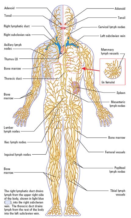
Lymphatic System
The lymphatic system is made up of lymphatic tissue, lymphatic vessels, and lymph fluid (Figure 4-10). This system is crucial in fighting infection and maintaining immune response to foreign substances. It also plays a role in draining excess fluid and transporting fat. T cells and B cells, which are important in providing an immune response to a foreign substance, are produced in lymphatic tissue. Lymphatic tissue is found in the bone marrow, thymus gland, lymph nodes, spleen, adenoids, and tonsils.
Lymph is a clear fluid that bathes cells. It flows through lymphatic vessels into the subclavian vein to rid the body of excess fluid.
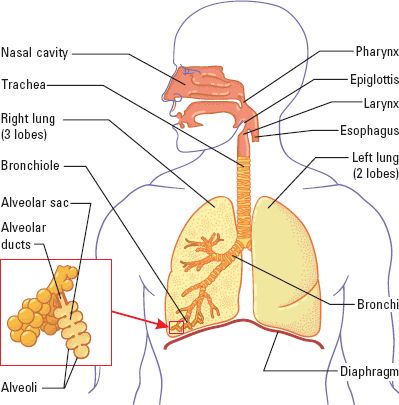
Respiratory System
The respiratory system includes the nasal cavity, pharynx (throat), larynx (voice box), trachea (windpipe), bronchi, and lungs (Figure 4-11). The primary function of this system is the exchange of gases that occurs between the alveoli and the capillaries.
Oxygen (O2) is absorbed into the blood from the air that has been breathed in. Carbon dioxide (CO2), a byproduct of cellular metabolism, is released from the blood and exhaled through the respiratory system.
If CO2 builds up, the blood becomes too acidic (the pH is too low), and this is toxic to cells. The pH value represents the hydrogen ion content of the blood. Similarly, if the pH of the blood is too high, the blood becomes too basic, and this, too, can damage cells throughout the body. Thus, the respiratory system is essential to maintaining the right concentration of hydrogen ions in the body. This is referred to as acid–base balance.
The respiratory system also permits the sense of smell, filters air that is breathed in, and produces sound (voice) as exhaled air interacts with the larynx in the throat.
Digestive System
The digestive, or gastroenterologic, system consists of the gastrointestinal tract, which runs from the mouth to the anus, and accessory digestive organs such as the pancreas, gallbladder, and liver (Figure 4-12). These structures have the following functions:
- Gastrointestinal tract—comprises the pharynx, esophagus, stomach, small intestine (duodenum, jejunum, and ileum), large intestine or colon, and rectum. Its job is to process food from the time it is eaten until it is digested, or broken down into small subunits, and then absorbed or eliminated. The degree of acidity changes along the gastrointestinal tract, and this affects the absorption and breakdown of food and drugs. In the stomach, the pH is low (very acidic), causing ingested substances to be broken down, but little absorption occurs for most nutrients and drugs. One notable exception is ethanol—the alcohol people consume in beverages—which is absorbed from the stomach. This accounts for its rapid onset of effects. Nutrients, water, and drugs are primarily absorbed in the small intestine, where the pH is higher (more basic).
- Pancreas—produces and secretes pancreatic enzymes that aid in breaking down food. The pancreas also produces hormones such as insulin and glucagon. These enzymes were mentioned above under the discussion of the endocrine system, and they are important in patients who have diabetes.
- Liver—detoxifies or breaks down ingested substances. The liver secretes bile, which is needed for fat absorption and is important in the breakdown or metabolism of carbohydrates and proteins. It also helps maintain the proper blood glucose level, stores some vitamins and minerals, and produces compounds that allow the blood to clot when needed. As shown in Figure 4-13, drugs are often metabolized (broken down or inactivated) in the liver by various hepatic (liver) enzymes (specialized proteins that facilitate chemical changes) and the cytochrome P450 (CYP 450) enzymes. These enzymes are involved in many drug interactions when two drugs are metabolized by the same enzyme, or when one drug alters the function of an enzyme responsible for metabolizing a second drug. Also, drugs absorbed from the intestines are carried directly to the liver, where they undergo first-pass metabolism. As shown in the lower part of Figure 4-13, some drugs are extensively metabolized at this point, meaning that only a small part of an oral dose actually reaches the general circulation of the body. This is called high, or extensive, first-pass metabolism. Drugs that undergo little transformation are said to undergo low first-pass metabolism. Metabolism is discussed further later in this chapter in the section on Pharmacokinetics.
- Gallbladder—stores bile, a yellowish-green substance that when secreted through bile ducts into the small intestine, helps the body emulsify fats so that they can be absorbed into the blood.