DNA Technology

Chapter 11 DNA Technology


Molecular genetics is the most active field in medical research today, and for good reason. Identification of the genetic risk factors for complex diseases allows us to understand the mechanisms of these diseases and to find targets for treatment. The diagnosis of genetic disease susceptibilities helps in disease prevention, diagnosis of diseases, and choice of treatments. Thus we see the emergence of a “personalized medicine” that is tailored to the patient’s genetic constitution. Although diagnosis is the main medical application of molecular genetics, genetic treatment modalities are being explored as well. This chapter introduces the basic toolkit that is used for these applications.


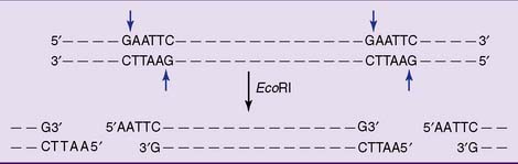
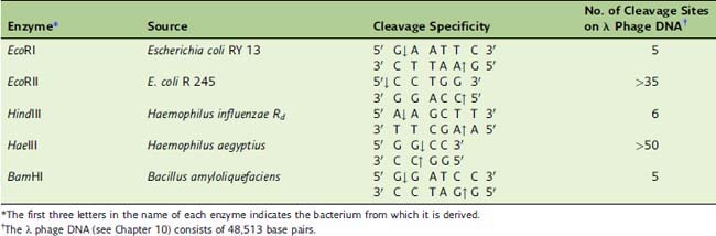
Restriction endonucleases cut large DNA molecules into smaller fragments


The DNA in human chromosomes has lengths of up to 300 million base pairs. For most applications, these unwieldy molecules need to be broken into smaller fragments.


The choice tools for DNA fragmentation are restriction endonucleases.These bacterial enzymes cleave DNA selectively at palindromic sequences of four to eight nucleotides. The average length of the resulting restriction fragments depends on the length of the recognition sequence. For example, an enzyme that recognizes a four-base sequence cleaves on average once every 256 (44) nucleotides and creates fragments with an average length of 256 base pairs. An enzyme that recognizes an eight-base sequence creates fragments with an average length of 65,536 (48) base pairs. Several hundred restriction endonucleases that recognize different palindromic sequences are available commercially.


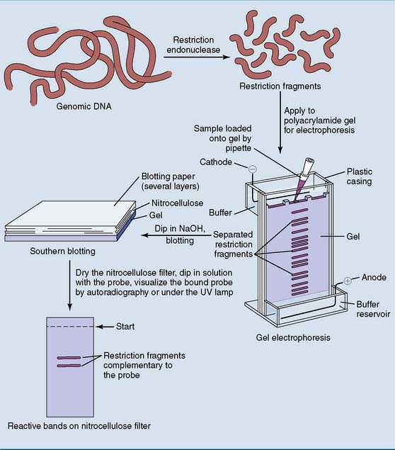
Most restriction enzymes make staggered cuts one or two base pairs away from the symmetry axis of their recognition sequence in both strands. Therefore the double-stranded restriction fragments have short single-stranded ends (Fig. 11.1 and Table 11.1).


Because the single-stranded overhangs are complementary to one another, every restriction fragment can anneal (base pair) with any other restriction fragment produced by the same enzyme. The annealed restriction fragments can be linked covalently by DNA ligase. This is the most fundamental procedure in recombinant DNA technology: the cutting and joining of DNA in the test tube.


Restriction endonucleases are produced by bacteria as a defense against DNA viruses. The bacteria protect susceptible sites in their own genome by methylation, but the unmethylated viral DNA is cut to pieces by the restriction enzyme.


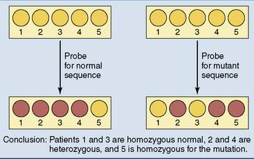
Dot blotting is used for genetic screening


The diagnosis of small mutations requires a synthetic oligonucleotide probe.The probe must be at least 17 or 18 nucleotides long because shorter probes are likely to hybridize with multiple sites in the genome. Oligonucleotides of this size can be synthesized by chemical methods.


The identification of a small mutation or polymorphism requires a pair of allele-specific oligonucleotides with identical lengths, one complementary to the normal sequence and the other complementary to the mutation. These probes are applied under conditions of high stringency. These are conditions of high temperature and/or low ionic strength that destabilize base pairing and permit annealing only if the sequences match precisely. Under conditions of low stringency, the probes would bind irrespective of the mismatch, and discrimination would be impossible.


Dot blotting (Fig. 11.2) is a rapid, inexpensive screening test for the detection of small mutations and polymorphisms. The extracted and denatured DNA is applied to two strips of nitrocellulose paper, which binds the single-stranded DNA tightly. One strip is dipped into a solution containing an oligonucleotide probe for the normal sequence, and the other is dipped into a solution with a probe for the mutation. If only the probe for the normal sequence binds, the patient is homozygous normal. If only the probe for the mutation binds, the patient is homozygous for the mutation. If both probes bind, the patient is heterozygous.


Cystic fibrosis (CF), for example, is a severe recessively inherited disease that affects 1 in 2500 newborns of European descent. About 4% of the population are heterozygous carriers of a CF mutation, and the risk of two carrier parents having an affected child is 25%.


In theory, CF is easy to prevent. All that is needed is to screen the whole population for CF mutations, identify all couples at risk, and persuade them to refrain from producing affected children. Other than a child-free lifestyle, the options for these couples include donor gametes, intrauterine or postnatal adoption, prenatal diagnosis with selective termination of affected pregnancies, and preimplantation genetic diagnosis with selective implantation of unaffected embryos.


More than 100 CF mutations are known, but a three–base-pair deletion (ΔPhe508 mutation) accounts for 50% to 70% of all CF mutations in the white population. For genetic screening, the DNA of large numbers of people is subjected to dot blotting with three to more than a dozen probes for the most common CF mutations in the local population. This design misses the rare mutations, but 80% to 90% of all CF carriers are identified.


Southern blotting determines the size of restriction fragments


Southern blotting (named after Ed Southern, who developed the method in 1975) provides information not only about the presence of a mutation but also about the length of the restriction fragment carrying the mutation.


As shown in Figure 11.3, restriction fragments obtained from genomic DNA are separated by electrophoresis in a cross-linked agarose or polyacrylamide gel. This method separates the restriction fragments by their size rather than their charge/mass ratio. Small fragments move fast, and large fragments move slowly because they are retarded by the gel.


The DNA is denatured by dipping the gel into a dilute sodium hydroxide solution, and then is transferred (“blotted”) to nitrocellulose paper to which it binds tightly. A replica of the gel with its separated restriction fragments is made on the nitrocellulose.


The desired fragment is identified by dipping the nitrocellulose paper in a neutral solution of the probe and washing off the excess unbound probe. Multiple fragments can be identified by using probes for different target sequences that are labeled with different fluorescent groups.


Northern blotting is a similar procedure for the analysis of RNA rather than DNA. Western blotting is a method for the separation of polypeptides that are then analyzed by monoclonal antibodies.


DNA can be amplified with the polymerase chain reaction


Southern blotting requires about 10 μg of DNA. This amount can be obtained from 1 ml of blood or from 10 mg of chorionic villus biopsy material. When less than this amount is available for analysis, the DNA has to be amplified with the polymerase chain reaction (PCR).


The procedure (shown in Figure 11.4) uses a heat-stable DNA polymerase such as Taq polymerase. This enzyme is derived from Thermus aquaticus, a thermophilic bacterium that was originally isolated from a hot spring in Yellowstone National Park. It functions best at temperatures close to 60°C and can survive repeated heating to 90°C.
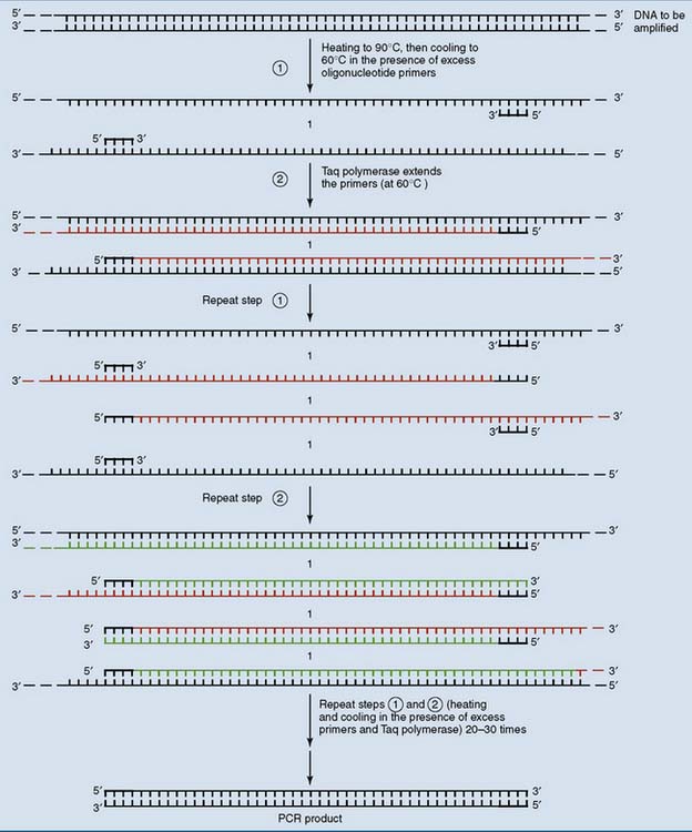


Like other DNA polymerases, Taq polymerase requires a primer. To amplify a defined section of genomic DNA, a pair of oligonucleotide primers that are complementary to the ends of the targeted DNA on both strands is used. The primers are added to the DNA in very large (>108-fold) molar excess, along with Taq polymerase and the precursors deoxy-ATP (dATP), deoxy-GTP (dGTP), deoxy-CTP (dCTP), and deoxy-TTP (dTTP). This mix is repeatedly heated to 90°C in order to denature the target DNA and cooled to 60°C for annealing of the primers and polymerization.


The target DNA is replicated in each cycle, and a single DNA molecule can be amplified to more than one million copies in about 1 hour. The resulting PCR product is a blunt-ended, double-stranded DNA that has the primers incorporated at its ends. It can be analyzed either by electrophoresis alone or by the use of allele-specific probes.


PCR has been used to amplify DNA from buccal smears, from single hairs sent to the laboratory in the mail or found at the scene of a crime, and even for sequencing of the Neanderthal genome from 40,000-year-old bones. However, it is difficult to reliably amplify DNA sequences longer than three kilobases. Individual exons can be amplified easily, but most genes are too large to be amplified in one piece. One limitation is that the Taq polymerase has no proofreading 3′-exonuclease activity. Therefore it misincorporates bases at a rate of about 1 every 5000 to 10,000 base pairs.


PCR is used for preimplantation genetic diagnosis


PCR is especially useful in prenatal diagnosis.The aim of prenatal diagnosis is the detection of severe fetal defects, with the option of terminating affected pregnancies. Fetal cells can be obtained by chorionic villus sampling at about 10 weeks of gestation or by amniocentesis at about 16 weeks. In this context, PCR is used to obviate the time-consuming culturing of fetal cells.


Preimplantation genetic diagnosis is a high-tech alternative to prenatal diagnosis. The embryo is produced by in vitro fertilization (IVF) and allowed to grow to the 8- or 16-cell stage. At this point, a single cell is removed from the embryo to supply the DNA for the diagnostic test. This does not impair further development of the embryo. Up to one dozen embryos are obtained in a single IVF cycle. All of them are subjected to the diagnostic test, and only the healthy ones are implanted.


PCR with nested primers is used to amplify DNA from a single cell. In this procedure, a section of the target DNA is amplified, and the amplification product is subjected to a second round of PCR with a more closely spaced primer pair.


Figure 11.5 shows the use of PCR with nested primers for preimplantation diagnosis of the ΔPhe508 mutation. This three–base-pair deletion is readily identified by PCR followed by gel electrophoresis because the mutated sequence yields a PCR product three base pairs shorter than normal. However, base substitutions cannot be identified by electrophoresis alone. They require the application of allele-specific probes to the PCR product.


PCR can detect deletions of entire exons or genes. For example, Duchenne muscular dystrophy is a fatal X-linked recessive muscle disease caused by deletions in the gene for the muscle protein dystrophin. With 79 exons scattered over more than two million base pairs of DNA, the dystrophin gene is the largest gene in the human genome. Most patients have large deletions that remove one or several exons from the gene.


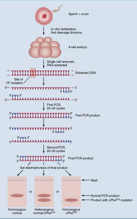
Figure 11.6 shows how these deletions are identified by amplification of deletion-prone exons. If one of the target exons is deleted, its PCR product is absent.
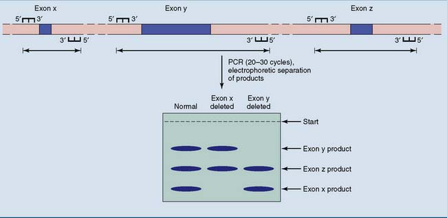


Allelic heterogeneity is the greatest challenge for molecular genetic diagnosis


All patients with sickle cell disease have the same mutation. Therefore a single pair of allele-specific oligonucleotide probes is sufficient for molecular diagnosis. However, this is an unusual situation. More commonly, any loss-of-function mutation that prevents the synthesis of a functional protein product will cause disease. In some diseases, such as CF, a small number of mutations accounts for a large majority of the cases. Therefore most carriers can be identified with a small assortment of oligonucleotide probes, one for each common mutation.


In the worst cases, most or all mutations for the disease are rare. In the X-linked clotting disorder hemophilia B, for example, more than 2000 different mutations in the gene for clotting factor IX have been observed in different patients. This degree of allelic heterogeneity makes the use of allele-specific oligonucleotide probes impractical. There are three ways to obviate this problem:


Normal polymorphisms are used as genetic markers


A polymorphism is defined as any DNA sequence variant for which the population frequency of the less common allele is more than 1%. Single-nucleotide polymorphisms (SNPs) are the most common type. There are more than 15 million SNPs in the human genome. They can be analyzed with allele-specific probes or with DNA microarrays.


Some SNPs obliterate or create a cleavage site for a restriction endonuclease. This subset of SNPs produces restriction-site polymorphisms (RSPs). They give rise to restriction fragments of different sizes that can be separated easily by gel electrophoresis.


Microsatellite polymorphisms are even more useful. These are tandemly repeated sequences with, in most cases, two to four nucleotides in the repeat unit and a total length well below 1000 base pairs (Fig. 11.7, B). The number of repeat units, and therefore the length of the microsatellite, varies among people. Whereas SNPs and RSPs have only two alleles, the most useful microsatellites have more than two alleles. Polymorphic microsatellites produce restriction fragments and PCR products of different length that can be separated by gel electrophoresis.
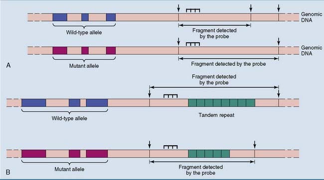


Most of these genetic markers do not cause disease. However, when a disease-causing mutation arises next to a polymorphic site on the chromosome, disease mutation and normal polymorphism travel together through the generations until they get divorced by a crossing-over in meiosis. Therefore the inheritance of the mutation can be traced by tracing the inheritance of the polymorphic markers with which it is associated.


Linkage patterns are different in different families. For example, the same mutation can arise next to a short variant of a neighboring microsatellite in one family and next to a long variant of the same microsatellite in another family. Therefore linkage cannot be used for population screening; it can be used only for studies of families in which the genotypes of one or more affected individuals are known.


Tandem repeats are used for DNA fingerprinting


Polymorphic DNA sequences can be used to identify criminals and, as has happened in many cases, for exonerating prisoners who had been wrongly convicted. This application is called DNA fingerprinting. Any polymorphism can be used, but microsatellite polymorphisms are most useful for DNA fingerprinting.


DNA fingerprinting can be performed with Southern blotting or PCR (Fig. 11.8). Southern blotting requires a substantial amount of DNA, for example, from a drop of seminal fluid from a sex offender. PCR is used when only a small amount of DNA is available, for example, from a single hair of the murderer stuck under the victim’s fingernail. However, because of its high sensitivity, PCR is more vulnerable to contamination by extraneous DNA. This could put the laboratory technician at risk for being wrongly convicted!
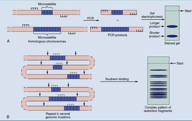


Another use of polymorphic microsatellites is paternity testing. Unlike the time-honored method of blood group typing, DNA tests allow an almost 100% accurate determination of paternity, unless the candidate fathers are monozygotic twins.


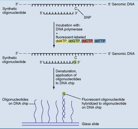
DNA microarrays can be used for genetic screening


Dot blotting tests for only one or a few mutations or polymorphisms at a time. DNA microarrays, also known as “DNA chips,” permit simultaneous testing of up to one million genetic variants.


An oligonucleotide microarray is prepared from a glass slide that is subdivided into up to one million little squares. Through photochemical methods, oligonucleotide probes with a length of 20 to 60 nucleotides are synthesized on each square. Each square receives a different oligonucleotide that is complementary to a short stretch of genomic DNA. Different probes are made for the alternative alleles of a polymorphic site.


Fluorescent-labeled genomic DNA fragments that are applied to the microarray hybridize primarily with the exact complementary probe but not the probe for the alternative allele. For example, when one square of the microarray has a probe for the sickle cell mutation and another has a probe for the corresponding normal sequence, normal DNA produces substantially more fluorescence on the square for the normal sequence. DNA from a sickle cell patient produces substantially more fluorescence on the square with the probe for the sickle cell mutation, and the DNA of a heterozygote produces about equal fluorescence on both. However, more imaginative methods are being explored as well (Fig. 11.9).


Jun 18, 2016 | Posted by in BIOCHEMISTRY | Comments Off on DNA Technology

Full access? Get Clinical Tree

Get Clinical Tree app for offline access