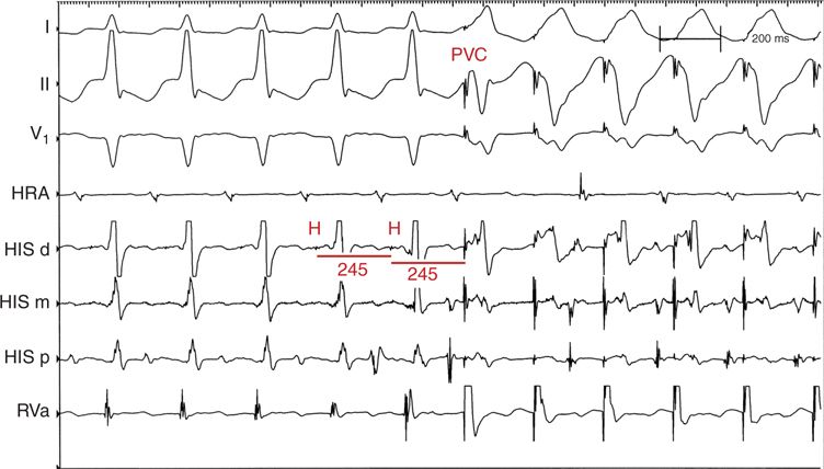
FIGURE 5-1 An example is shown of a PVC delivered during His bundle refractoriness during PSVT in the EP laboratory. There is preexcitation of the atrium with a His-synchronous PVC, which is evidence of an accessory pathway but is not diagnostic of AVRT. Although unlikely, an atrial tachycardia with a concealed bystander accessory pathway cannot be excluded. The His bundle is refractory as seen by the coincident antegrade His potential. Thus, retrograde conduction of the PVC must have occurred over an accessory pathway.
• Advancement of atrial activation with a PVC delivered during PSVT when the His bundle should be refractory as seen in this patient indicates that a retrogradely conducting AP is present, but it may not necessarily participate in tachycardia. Although unlikely, an atrial tachycardia with a concealed bystander AP cannot be excluded.
• A His-synchronous PVC that terminates the tachycardia without preexcitation of the atrium is diagnostic of AVRT (Figure 5-2). An atrial tachycardia would not terminate under those conditions. Note that in this example there was an attempt to entrain the atrium with ventricular pacing, but pacing terminated tachycardia. Careful examination showed that the first beat of the pacing train occurred during the His bundle refractory period and ultimately provided the diagnosis of AVRT.
FIGURE 5-2 Termination of tachycardia with a His-synchronous PVC without preexcitation of the atrium is shown. This is diagnostic of AVRT.
• The Principle: A PVC that preexcites the atrium must do so via an AP because when the His-Purkinje system (HPS) is refractory during PSVT, a PVC cannot conduct to the atrium through the HPS. Similarly, a PVC that terminates tachycardia without preexciting the atrium must have occurred as a result of causing block in the AP. When atrial activation is delayed with a PVC delivered during His refractoriness, the maneuver is also diagnostic for AVRT because an AP must be participating in the tachycardia.
• Pitfall: Preexcitation of the atrium may not occur despite the presence of an AP if the pacing site is far from the ventricular insertion of the AP; the ability of the ventricular extrastimulus to enter the reentrant circuit before ventricular activation over the normal pathway is affected by (1) the conduction time from the ventricular stimulation site to the AP, (2) the local ventricular refractory period, and (3) the tachycardia cycle length.
Keys to Performing This Maneuver
• Scan diastole with PVCs at progressively shorter coupling intervals (decrease by 10 ms).
• Confirm ventricular capture.
Stay updated, free articles. Join our Telegram channel

Full access? Get Clinical Tree


