Caution
In surgery ROCM are often referred to incorrectly as “dyes.” Do not become confused! ROCM are an entirely different category of diagnostic agents used for very different purposes.
Dyes are solutions that color or mark tissue for identification. Dyes may be used to mark skin incisions, delineate normal tissue planes, or enhance visualization of certain anatomic structures during a surgical procedure. Dyes may be applied topically, injected into the bloodstream, or instilled into a body cavity.
Staining agents are used in surgery to help visually identify abnormal cells, most frequently in procedures on the cervix. Staining agents are chemicals in solution that react differently with abnormal cells from the way they react with normal cells.
Contrast Media
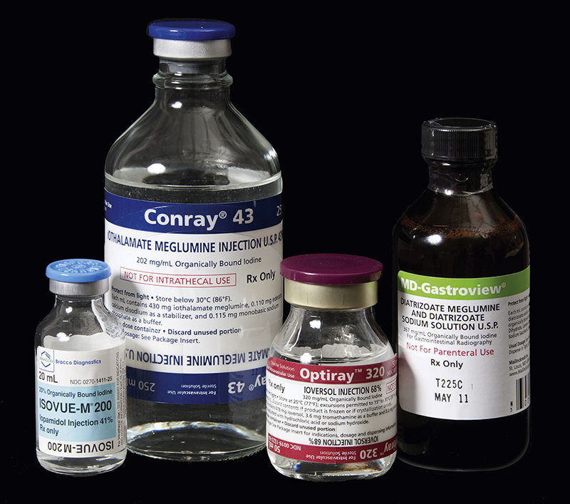
Contrast media are high-density pharmacological agents (Fig. 6-1) used to visualize low-contrast body tissues that include vascular structures, the urinary bladder, kidneys, the gastrointestinal (GI) tract, and the biliary tree. Although these agents are primarily used in the diagnostic imaging department of health care facilities, some diagnostic imaging procedures are performed in surgery. Many different contrast media are available for various diagnostic examinations, several of which are provided in Table 6-1 as examples. ROCM are classified into two main categories: ionic (molecules that break apart into ions when dissolved in water) and nonionic (do not break apart when dissolved in water).
All contrast media used in surgery contain iodine; therefore a thorough patient history of allergies or reactions to iodine must be obtained and noted in the chart (this includes shellfish allergies). The circulator will also check for a history of patient allergies or reactions to iodine during the preoperative assessment.
 Caution
If the patient has a sensitivity to iodine, a facility may require a premedication protocol of prednisone and diphenhydramine (Benadryl) before using any ROCM containing iodine.
If the patient has a positive history for iodine reaction and use of contrast media is anticipated during the surgical procedure, the anesthesia care provider and the surgeon should be alerted before patient transport to the operating room. The surgical technologist should prepare for these patients by having selection of nonionic contrast agents available in the operating room.
Most reactions to ROCM are mild and limited, requiring little or no treatment. However, severe allergic reactions can result in a life-threatening condition called anaphylaxis (see Chapter 16), which requires a rapid and definitive treatment protocol.
The use of ROCM provides an excellent example of pharmacokinetics in action.
 Quick Question
What does the term “pharmacokinetics” mean, and what are the four processes involved in pharmacokinetics? See Chapter 1 to check your answers.

FIGURE 6-1 Various iodinated contrast agents. (From Adler AM, Carlton RR: Introduction to radiologic and imaging sciences and patient care, ed 6, St Louis, 2016, Elsevier.)
TABLE 6-1
Examples of Radiopaque Contrast Media
| Trade Name | Generic Name |
| Hypaque-76 | diatrizoate meglumine 66% and diatrizoate sodium 10% |
| Hypaque-Cysto | diatrizoate meglumine |
| Cystografin | diatrizoate meglumine |
| Renografin-60 | diatrizoate meglumine and diatrizoate sodium |
| Conray 43 | iothalamate meglumine |
| Omnipaque | Iohexol |
| Isovue | iopamidol |
| Cholografin | iodipamide meglumine |
| Optiray | ioversol |
| Ultravist | iopromide |
| Visipaque | iodixanol |
When administered intravenously, a contrast agent is absorbed immediately because it directly enters the circulatory system for distribution. The agent is diluted by the circulating volume of blood as it continues to be distributed. Most ROCM are not highly bound to circulating plasma proteins and do not undergo significant biotransformation in the liver. Thus ROCM molecules are excreted virtually unchanged through renal glomerular filtration. The presence of active ROCM in the urinary system enables visualization of the kidneys, ureters, and bladder with intravenous urography (IVU). Intra-arterial administration of ROCM (e.g., as seen in surgery during placement of aortic endostent grafts or femoral embolectomy) enables immediate visualization of intended blood vessels on an arteriogram.
< div class='tao-gold-member'>
Only gold members can continue reading. Log In or Register to continue


