201 Diabetic retinopathy
Instruction
Examine the fundus in these patients.
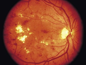
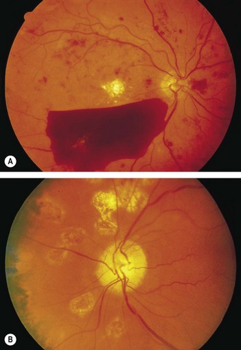
• You will be expected to comment on whether it is background (Fig. 201.1) or proliferative (Fig. 201.2) retinopathy.
• You may have a clue about the underlying diabetes, either from a diabetic chart or from the presence of diabetic fruit juices at the bedside.
Patient 1
Salient features
Examination


Fig. 201.3 Diabetic eye disease. (A) Normal macula and optic disc. (B) Early background retinopathy, dot and blot haemorrhages. (C) Early background retinopathy, plus hard exudates. (D) Preproliferative retinopathy, with multiple cotton-wool spots. (E) Proliferative retinopathy, with hallmark frond-like new vesicles. (F) Exudative maculopathy, with exudates within a disc-width of the macula.(G,H) Central (G) and cortical (H) cataracts seen against the red reflex of the ophthalmoscope.
(With permission from Kumar, Clark 2005.)
Diagnosis
This patient has dot and blot haemorrhages, and cottonwool spots (lesions), probably caused by diabetic retinopathy (aetiology), and good visual acuity (functional status).
Questions
How would you manage such a patient?
The Early Treatment of Diabetic Retinopathy Study (ETDRS) has established that early peripheral (panretinal) argon laser photocoagulation is not indicated for mild to moderate non-proliferative retinopathy. Early treatment in the form of argon laser photocoagulation applied directly to leaking microaneurysms, as well as grid photocoagulation applied to diffuse areas of leakage and thickening, is highly beneficial.

Full access? Get Clinical Tree

