Diabetes mellitus

44 Diabetes mellitus


Diabetes mellitus is the most common of the endocrine disorders. It is a chronic condition, characterised by hyperglycaemia and due to impaired insulin secretion with or without insulin resistance. Diabetes mellitus may be classified according to aetiology, by far the most common types being type 1 and type 2 diabetes (Box 44.1). More than 2.6 million people in the UK have diabetes, and by the year 2025, this number is estimated to rise to 4 million.


Type 1 diabetes is a disease characterised by the destruction of the insulin-producing pancreatic β-cells, the development of which is either autoimmune T-cell mediated destruction (type 1A) or idiopathic (type 1B). In over 90% of cases, β-cell destruction is associated with autoimmune disease. Type 1 diabetes usually develops in the young (below the age of 30), although it can develop at any age and is usually associated with a faster onset of symptoms leading to dependency on extrinsic insulin for survival.


Type 2 diabetes is more common above the age of 40, with a peak age of onset in developed countries between 60 and 70 years, although it is being increasingly seen in younger people and even children. The prevalence of type 2 diabetes varies widely in different populations, being six times more common in those of South Asian origin compared with those of Northern European origin. It is caused by a relative insulin deficiency and insulin resistance. Symptoms are generally slower in onset and less marked than those of type 1. Type 2 diabetes may be an incidental finding, particularly when patients present with complications associated with the disease, for example, heart disease. Type 2 disease often progresses to the extent whereby extrinsic insulin is required to maintain blood glucose levels. The differences between type 1 and type 2 diabetes are highlighted in Table 44.1. It is sometimes difficult to distinguish clinically between type 1 and type 2 diabetes. The important thing to be aware of is that it is predominantly the degree of metabolic abnormality that is the key determinant of the form of treatment.


Table 44.1 Differences between type 1 and type 2 diabetes


Type 1 diabetes Type 2 diabetes
β-cell destruction No β-cell destruction
Islet cell antibodies present No islet cell antibodies present
Strong genetic link Very strong genetic link
Age of onset usually below 30 Age of onset usually above 40
Faster onset of symptoms Slower onset of symptoms
Insulin must be administered Diet control and oral hypoglycaemic agents often sufficient control
Patients usually not overweight Patients usually overweight
Extreme hyperglycaemia causes diabetic ketoacidosis Extreme hyperglycaemia causes hyperosmolar hyperglycaemic state

Two other varieties of non-typical diabetes that may be seen are latent autoimmune diabetes in adults (LADA) and maturity-onset diabetes of the young (MODY). LADA occurs in younger, leaner individuals who appear to have type 2 diabetes as they do not become ketotic and may manage without insulin for a time. Antiglutamic acid decarboxylase (GAD) antibodies may be present and the individual usually progresses to insulin more rapidly than those with other varieties of type 2 diabetes. MODY was noted over 30 years ago and described a subset of type 2 diabetes of young onset, often with a positive family history. Genetic studies have now identified this to be a monogenic autosomal dominant form of diabetes. MODY related to the glucokinase gene typically causes a resetting of the glucose level with a ‘mild’ non-progressive hyperglycaemia in which diet treatment is usually sufficient. Other types of MODY are related to mutations in the hepatocyte nuclear factor genes and usually develop during adolescence or the early 20s. Pharmacological treatment is required, but sulphonylureas are extremely effective and insulin can usually be avoided.


Epidemiology


The incidence of type 1 diabetes is increasing worldwide, for unknown reasons. It is speculated that environmental changes may be causing modification to the diabetes-associated alleles. Also, since the introduction of insulin in the 1930s, an increasing number of people with type 1 diabetes have had children. There are major ethnic and geographical differences in the prevalence and incidence of type 1 diabetes. Figures are highest in Caucasians (especially Scandinavians), while the disorder is rare in Japan and the Pacific area. In northern Europe, the prevalence is approximately 0.3% in those under 30 years of age. Type 1 diabetes may present at any age, but there is a sharp increase around the time of puberty and a decline thereafter. Approximately 50–60% of patients with type 1 will present before 20 years of age.


Type 2 diabetes is much more common than type 1, accounting for 90% of people with diabetes. It usually occurs in those over the age of 40 years. Estimates in the UK suggest that type 2 diabetes currently affects approximately 2.3 million people, and up to another 500,000 are thought to be undiagnosed. The incidence of type 2 rises with age and with increasing obesity. As with type 1, there are major ethnic and geographical variations. In general, in non-obese populations, the prevalence is 1–3%. In the more obese societies, there is a sharp increase in prevalence with estimates of 6–8% in the USA, increasing to values as high as 50% in the Pima Indians of Arizona. Diabetes is six times more common among Asian immigrants in the UK than in the indigenous population. World studies of immigrants have suggested that the chances of developing type 2 are between two and 20 times higher in well-fed populations than in lean populations of the same race.


Aetiology


Both genetic and environmental factors are relevant in the development of type 1 diabetes, but the exact relationship between the two is still unknown. There is a strong immunological component to type 1 and a clear association with many organ-specific autoimmune diseases. Circulating islet cell antibodies (ICAs) are present in more than 70% of those with type 1 at the time of diagnosis. Family studies have shown that the appearance of ICAs often precedes the onset of clinical diabetes by as much as 3 years. Type 1 has been widely believed to be a disease of clinically rapid onset, but the development is related to a slow process of progressive immunological damage. However, it is not currently possible to use screening methods to reliably identify patients who will develop diabetes in the future. The final event that precipitates clinical diabetes may be caused by sudden stress such as an infection when the mass of β-cells in the pancreas falls below 5–10%.


Studies have been carried out in which patients with newly diagnosed type 1 were treated with immunosuppressive therapies such as ciclosporin, azathioprine, prednisolone and antithymocyte globulin. When started soon after diagnosis, these therapies showed transient improvements in clinical measures and increased the rate of remissions in which insulin was not required. However, their use is limited in an otherwise healthy and young population due to potential toxicity and the risks associated with immune suppression.


Studies have investigated the use of anti-CD3 monoclonal antibodies. When newly diagnosed type 1 patients are treated with short courses of anti-CD3 monoclonal antibodies, smaller insulin doses are required. This relates to better preservation of β-cell function.


Type 2 diabetes also has a strong genetic predisposition. Identical twins have a concordance rate approaching 100%, suggesting the relative importance of inheritance over environment. If a parent has type 2, the risk of a child eventually developing type 2 is 5–10% compared with 1–2% for type 1. Type 2 diabetes occurs because of the progressive development of insulin resistance and β-cell dysfunction, the latter leading to an inability of the pancreas to produce enough insulin to overcome the insulin resistance. About 85% of people with type 2 diabetes are obese. This highlights the clear association between type 2 and obesity, with obesity causing insulin resistance. In particular, central obesity, where adipose tissue is deposited intra-abdominally rather than subcutaneously, is associated with the highest risk. Body mass index (BMI) has been used as an indicator for predicting type 2 risk; however, it does not take fat distribution into account, so waist circumference measurements are now being increasingly used.


Pathophysiology


The islets of Langerhans form the endocrine component of the pancreas, constituting 1% of the total pancreatic mass. Insulin is synthesised in the pancreatic β-cells, initially as a polypeptide precursor, preproinsulin. The latter is rapidly converted in the pancreas to proinsulin. This forms equal amounts of insulin and C-peptide through removal of four amino acid residues. Insulin consists of 51 amino acids in two chains (the A chain contains 21 amino acids and B chain contains 30), connected by two disulphide bridges. In the islets, insulin and C-peptide (and some proinsulin) are packaged into granules. Insulin associates spontaneously into a hexamer containing two zinc ions and one calcium ion.


Glucose is the major stimulant to insulin release. The response is triggered both by the intake of nutrients and the release of gastro-intestinal peptide hormones. Following an intravenous injection of glucose, there is a biphasic insulin response. There is an initial rapid response in the first 2 min, followed after 5–10 min by a second response which is smaller but sustained over 1 h. The initial response represents the release of stored insulin and the second phase reflects discharge of newly synthesised insulin. Glucose is unique; other agents, including sulphonylureas, do not result in insulin biosynthesis, only release. Once released from the pancreas, insulin enters the portal circulation. The liver rapidly degrades it and only 50% reaches the peripheral circulation. In the basal state, insulin secretion is at a rate of approximately 1 unit/h. The intake of food results in a prompt five- to tenfold increase. Total daily secretion is approximately 40 units.


Insulin circulates free as a monomer, has a half-life of 3–5 min and is primarily metabolised by the liver and kidneys. In the kidneys, insulin is filtered by the glomeruli and reabsorbed by the tubules and degraded. In both renal and hepatic disease, there is a decrease in the rate of insulin clearance, which may necessitate dosage reduction for those using exogenous insulin. Peripheral tissues such as muscle and fat also degrade insulin, but this is of minor quantitative significance.


The interaction of insulin with the receptor on the cell surface sets off a chain of messengers within the cell. This opens up transport processes for glucose, amino acids and electrolytes.


In type 1 diabetes, there is an acute deficiency of insulin that leads to unrestrained hepatic glycogenolysis and gluconeogenesis with a consequent increase in hepatic glucose output. Also, glucose uptake is decreased in insulin-sensitive tissues such as adipose tissue and muscle; hence, hyperglycaemia ensues. Either as a result of the metabolic disturbance itself or secondary to infection or other acute illness, there is increased secretion of the counter-regulatory hormones glucagon, cortisol, catecholamine and growth hormone. All of these will further increase hepatic glucose production.


In type 2 diabetes, the process is usually less acute, since insulin production decreases over a sustained period of time. Hyperinsulinaemia is able to maintain glucose levels for a period of time, but eventually β-cell function deteriorates and hyperglycaemia ensues. If this cycle is not interrupted, type 2 diabetes develops. Impaired glucose tolerance (IGT), impaired fasting glucose or hyperinsulinaemia may be detected before overt diabetes develops, and if so, a strict diet and exercise regimen leading to weight loss and improved insulin sensitivity may delay or even prevent the onset of diabetes. At the time of diagnosis, those with type 2 diabetes may have already lost about 50% of their β-cell function. Irrespective of treatment, β-cell function continues to decline with time, often leading to the need for regular insulin therapy.


Type 2 diabetes is also associated with the metabolic syndrome (or syndrome X), although the real relevance of this ‘syndrome’ continues to be debated in the literature (Khan et al., 2005). The metabolic syndrome is a group of risk factors commonly found in those with type 2 diabetes, including insulin resistance, glucose intolerance (type 2 diabetes or IGT), hyperinsulinaemia, hypertension, dyslipidaemia, central obesity, atherosclerosis and increased levels of procoagulant factors, for example, plasminogen activator inhibitor-1 and fibrinogen.


Pathophysiology of insulin resistance


Abdominal fat, found in abundance in the majority of those with type 2 diabetes, is metabolically different from subcutaneous fat and can cause ‘lipotoxicity’. Abdominal fat is resistant to the antilipolytic effects of insulin, resulting in the release of excessive amounts of free fatty acids, which in turn lead to insulin resistance in the liver and muscle. The effect is an increase in gluconeogenesis in the liver and an inhibition of insulin-mediated glucose uptake in the muscle. Both these result in increased levels of circulating glucose. Further, excess fat itself may contribute to insulin resistance because when adipocytes become too large they are unable to store additional fat, resulting in fat storage in the muscles, liver and pancreas, causing insulin resistance in these organs.


Excess intra-cavity adipose tissue causes the oversecretion of some cytokines (adipokines or adipocytokines) associated with inflammation, endothelial dysfunction and thrombosis. Examples of such adipokines include plasminogen activator inhibitor-1 (which is prothrombotic), tumour necrosis factor-α and interleukin-6 (which are proinflammatory) and resistin (which causes insulin resistance). The atherosclerosis associated with insulin resistance is due to hypercoagulability, impaired fibrinolysis and the toxic combination of endothelial damage (caused by chronic, subclinical inflammation), oxidative stress and hyperglycaemia. Excess adipose tissue is also thought to cause undersecretion of a beneficial adipokine called adiponectin. Adiponectin suppresses the attachment of monocytes to endothelial cells, thereby protecting against vascular damage. People with type 2 diabetes have lower levels of adiponectin than those without diabetes and weight reduction increases adiponectin levels.


Clinical manifestations


The symptoms of both type 1 and type 2 diabetes are similar, but they usually vary in intensity. Those associated with type 1 diabetes are more severe and faster in onset. The symptoms are related to the osmotic effects of glucose and the abnormalities of energy partitioning. Common symptoms include polyuria (increased urine production, particularly noticeable at night) and polydipsia (increased thirst). These are a consequence of osmotic diuresis secondary to hyperglycaemia. These symptoms are frequently accompanied by fatigue due to an inability to utilise glucose and marked weight loss because of the breakdown of body protein and fat as an alternative energy source to glucose. Blurred vision caused by a change in lens refraction may also occur and patients should be advised that as glucose levels are normalised, vision normally improves and new spectacles should be avoided for the first 3 months of effective treatment of the hyperglycaemia. Patients may also experience a higher infection rate, especially Candida, and urinary tract infections due to increased urinary glucose levels.


Diagnosis


In June 2000, the UK formally adopted the World Health Organization criteria for diagnosing diabetes mellitus that was initially published in 1999. It has since been updated and the diagnostic criteria have been reiterated (World Health Organization, 2006).


Current recommendations are that the diagnosis is confirmed by a glucose measurement performed in an accredited laboratory on a venous serum sample. A diagnosis should never be made on the basis of glycosuria or a stick reading of a finger prick blood glucose alone, although such tests are being examined for screening purposes. Glycated haemoglobin (HbA1c) is also not currently recommended for diagnostic purposes, although this is currently being considered.


Diabetic emergencies


Hypoglycaemia and extreme hyperglycaemia, causing diabetic ketoacidosis or hyperosmolar hyperglycaemic state, constitute the three acute emergencies associated with diabetes.


Hypoglycaemia


Hypoglycaemia can occur both with insulin treatment and in those taking some oral agents, especially the longer-acting sulphonylureas, for example, chlorpropamide and glibenclamide. Definitions of hypoglycaemia vary, and in particular, there is no WHO definition. However, symptoms caused by the release of counter-regulatory hormones predominantly adrenaline (epinephrine), noradrenaline (norepinephrine) and glucagon tend to occur when the venous serum glucose drops below 3.0 mmol/L in healthy individuals. These symptoms described in Box 44.2 are a normal physiological response to hypoglycaemia and should alert the person to consume carbohydrates. Individuals may not respond appropriately to hypoglycaemia of this degree for several reasons, termed hypoglycaemia unawareness. First, the relevance of the symptoms has not been explained to them. This is an educational failing. It is imperative, therefore, that people with diabetes who are prescribed medication which is known to cause hypoglycaemia should be educated about the autonomic symptoms so that they may take action to avoid further decline of serum glucose. Second, the symptoms simply may not occur because of autonomic neuropathy. One of the commonest complications of diabetes is neuropathy, and when this includes the autonomic nervous system, there are no reliable symptoms to warn the individual that they are hypoglycaemic. A similar situation may occur as a consequence of drugs which suppress autonomic symptoms, such as β-blockers. Third, the patient may have recurrent hypoglycaemia. In those individuals who suffer frequent hypoglycaemic episodes, the autonomic symptoms may cease to occur. There is some evidence that the symptoms can be regained if, for a period of a few weeks, the serum glucose level can be maintained out of the hypoglycaemic range. Finally, the individual may be hypoglycaemia unaware because of alcohol intoxication.


If the serum glucose is allowed to drop to around 2 mmol/L, there are acute changes in cerebral function which lead initially to confusion. This is followed by coma, seizures and death if the glucose drops below about 0.5 mmol/L. Any cerebral malfunction is termed neuroglycopaenia.


Nocturnal hypoglycaemia


Sometimes, hypoglycaemia occurs throughout the night. Symptoms may include restlessness, although this may not be identified unless observed by another person. When nocturnal hypoglycaemia occurs, the person often wakes feeling unrested, unwell or with a headache. Contrary to what might be expected, morning blood glucose readings may be high because a sustained hypoglycaemic episode leads counter-regulatory hormones to raise blood glucose levels. This could present a confusing picture as the obvious solution to a raised blood glucose level in the morning would be to increase the evening/night-time dose of insulin. However, in the case of nocturnal hypoglycaemia, this would make the problem worse. If nocturnal hypoglycaemia is suspected, then blood glucose should be measured at night, for example, 2.00–3.00 am. If confirmed, the patient should either have a snack before bedtime, reduce the evening/night-time dose of insulin, alter the timing of administration of the evening dose of intermediate- or long-acting insulin in order to delay the peak of bioavailability or change the intermediate-acting insulin to a peakless analogue as appropriate. It is important to discuss nocturnal hypoglycaemia with patients as there is often a fear of dying from unrecognised hypoglycaemia in sleep. This fear is unfounded because of the protection from hypoglycaemia severe enough to cause death afforded by the counter-regulatory hormones. This occurs even in those individuals with autonomic neuropathy.


Treatment of hypoglycaemia


If the patient is able to swallow safely without the risk of aspiration, then glucose should be taken orally. However, if unable to swallow or if there is a risk of aspiration because, for example, of a decreased level of consciousness, parenteral treatment should be given, either intravenous glucose or intramuscular glucagon.


The most effective oral treatments are pure sources of glucose, for example, five glucose tablets or glucose drinks such as 150 mL of Lucozade®. In an emergency, hot drinks should be avoided as they might burn and drinks containing milk are not suitable as the fat in milk slows down sugar absorption. Blood glucose levels should be measured about 10–15 min after treating hypoglycaemia. If below 3.5 mmol/L, more glucose should be consumed. If above 3.5 mmol/L and the next meal will be over 1 h, then a long-acting carbohydrate is also required, for example, bread or biscuits. However, if the person is taking an α-glucosidase inhibitor such as acarbose, then monosaccharide carbohydrates must be given because disaccharides and polysaccharides will not be absorbed due to inhibition of the enzymes cleaving carbohydrate into absorbable monosaccharide units.


Should parenteral treatment be required, 25 g of intravenous glucose or 1 mg of intramuscular glucagon is recommended. Glucagon takes approximately 15–20 min to work, but if the person has liver disease (cirrhosis) or is malnourished, then glucagon may not work because glucagon acts by mobilising glucose stores from the liver. In such cases, intravenous glucose must be given. A number of serious extravasation injuries, some necessitating amputation of the affected limb, have been caused by 50% glucose. As a consequence, many hospitals now use 20% glucose.


Diabetic ketoacidosis


Diabetic ketoacidosis is serious, and in developed countries, it has a mortality rate of 5–10%. It occurs because absence of insulin causes extreme hyperglycaemia. At the same time, the normal restraining effect of insulin on lipolysis is removed. Non-esterified fatty acids are released into the circulation and taken up by the liver, which produces acetyl coenzyme A (acetyl CoA). The capacity of the tricarboxylic acid cycle to metabolise acetyl CoA is rapidly exceeded. Ketone bodies, acetoacetate and hydroxybutyrate are formed in increased amounts and released into the circulation. Further, osmotic diuresis, caused by hyperglycaemia, lowers serum volume, causing dizziness and weakness due to postural hypotension. Weakness is increased by potassium loss, caused by urinary excretion and vomiting due to stimulation of the vomiting centre by ketones, and catabolism of muscle protein. When insulin deficiency is severe and of acute onset, all of these symptoms are accelerated. Ketoacidosis exacerbates the dehydration and hyperosmolarity by producing anorexia, nausea and vomiting. As serum osmolarity rises, impaired consciousness ensues with coma developing in approximately 10% of cases. Metabolic acidosis causes stimulation of the medullary respiratory centre, giving rise to Kussmaul respiration (deep and rapid breathing) in an attempt to correct the acidosis. The patient’s breath may have the fruity odour of acetone (ketones) commonly described as smelling like pear drops or nail varnish remover.


Precipitating factors for diabetic ketoacidosis in type 1 disease are usually omission of insulin dose, acute infection, trauma or myocardial infarction. Although diabetic ketoacidosis is normally associated with type 1 diabetes, it may rarely occur in people with type 2.


Hyperosmolar hyperglycaemic state


HHS is associated with type 2 disease and has a higher mortality rate (15%) than diabetic ketoacidosis. HHS usually occurs in middle-aged or elderly people, about 25% of whom have previously undiagnosed type 2 diabetes.


In HHS, unlike diabetic ketoacidosis, there is no significant ketone production and therefore no severe acidosis. Hyperglycaemia occurs gradually over a sustained period of time, leading to dehydration due to osmotic diuresis which, if severe, results in hyperosmolarity. Hyperosmolarity may increase blood viscosity and the risk of thromboembolism. Factors precipitating HHS are infection, myocardial infarction, poor adherence with medication regimens or medicines which cause diuresis or impair glucose tolerance, for example, glucocorticoids.


Long-term diabetic complications


Diabetes and its long-term complications cost the NHS substantial amounts of money – approximately 10% of the total budget (£173 million/week).


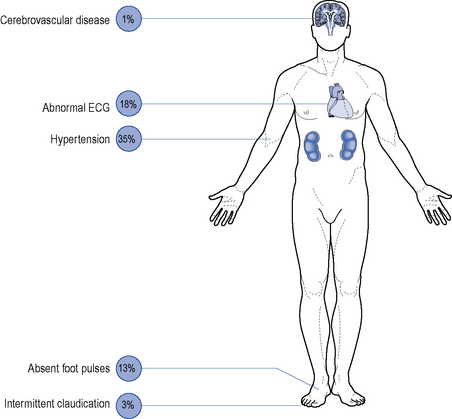
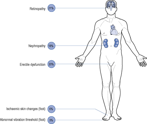
Although all long-term complications may occur in each type of diabetes, the spectrum of incidence is different. Many patients with type 2 diabetes have had their disease a long time before the diagnosis, by which time many have developed diabetic complications (Figs. 44.1 and 44.2). However, diabetic complications can be limited and sometimes prevented altogether if good management occurs from an early stage. Hyperglycaemia and hypertension are the two major modifiable risk factors that influence the development of diabetic complications.


Patients with diabetes should undergo regular review of their disease management for early signs of associated complications and review of these risk factors and their management. Diabetic complications are frequently divided into macrovascular and microvascular complications. Macrovascular complications arise from damage to large blood vessels and microvascular complications occur from damage to smaller vessels. The general aetiology of macro- and microvascular complications is the same and results from atherosclerosis of the vessels, which may lead to occlusion. The main aims of treatment are, first, to prevent the immediate symptoms associated with diabetes, for example, polyuria, polydipsia, etc., and second, to prevent development, or slow the progression of the long-term disease complications.


Macrovascular disease


The risk of macrovascular complications, including cardiovascular disease (coronary heart disease and stroke) and PVD, is 2–4 times higher for people with diabetes.


Cardiovascular disease


The most common cause of death in people with type 2 diabetes is cardiovascular disease which accounts for an estimated 80% of deaths in this patient group. The risk of a person with diabetes having a myocardial infarction (MI) is the same as someone without diabetes having a second myocardial infarction. The risk of cardiovascular disease is increased further if nephropathy is present. Other cardiovascular disease risk factors are the same as in the non-diabetic population and include smoking, hypertension and dyslipidaemia. However, these risk factors are enhanced in the presence of diabetes, and therefore smokers are encouraged to stop, and individuals with hypertension and lipid disorders are actively reviewed and treated. Silent myocardial infarction (infarction with no symptoms) is more common in those with diabetes and may be due to cardiac autonomic neuropathy. Cerebrovascular disease is also more commonly associated with diabetes, and patients have a greater mortality and morbidity compared to the general population.


Microvascular disease


Microvascular complications include retinopathy, nephropathy and neuropathy.


Nephropathy


In diabetic renal disease, the kidneys become enlarged and the glomerular filtration rate (GFR) initially increases. However, if the nephropathy progresses, the GFR starts to decline. Serum creatinine used alone to estimate renal function has limitations. The GFR can be estimated (eGFR). The most popular method is the modified Modification of Diet in Renal Disease (MDRD) formula, which requires serum creatinine, age, sex and ethnicity:


image


The presence of nephropathy is indicated by the detection of microalbuminuria (small amounts of albumin present in urine). If higher amounts of albumin are detected, this is termed proteinuria (or macroalbuminuria) and signifies more severe renal damage. Microalbuminuria is defined as an albumin:creatinine ratio (ACR) greater or equal to 2.5 mg/mmol (men) and 3.5 mg/mmol (women). Proteinuria may be defined as an albumin:creatinine ratio greater than 30 mg/mmol or albumin concentration greater than 200 mg/L. Proteinuria may progress to end-stage renal disease and require dialysis. Albumin in the urine increases the risk of cardiovascular disease, with microalbuminuria associated with 2–4 times the risk, proteinuria with nine times the risk and end-stage renal disease increasing risk by 50 times.


Tight control of both glycaemic levels and blood pressure reduces the risk of developing nephropathy. Angiotensin-converting enzyme (ACE) inhibitors and/or angiotensin receptor blockers (ARBs) are the treatments of choice, since both have been demonstrated to provide renal protective effects additional to their antihypertensive effects. ACE inhibitors and ARBs have been shown to delay the progression to proteinuria in patients with microalbuminuria. Although not proven for all individual drugs in these classes, it is considered to be a class effect. However, these drugs should be used with care if there is a risk of renovascular disease.


Peripheral neuropathy


Peripheral neuropathy is the progressive loss of peripheral nerve fibres resulting in nerve dysfunction. Diabetic neuropathies can lead to a wide variety of sensory, motor and autonomic symptoms. The most common is the symmetrical distal sensory type, which is particularly evident in the feet and may slowly progress to a complete loss of feeling. It is most prevalent in elderly patients with type 2 diabetes but may be found with any type of diabetes, at any age beyond childhood. Painful diabetic neuropathy is another manifestation of sensory neuropathy; it can be extremely disabling and may cause considerable morbidity. Guidance on the treatment of painful neuropathy is available (National Institute for Health and Clinical Excellence, 2010). Diabetic proximal motor neuropathy is rapid in onset and involves weakness and wasting, principally of the thigh muscles. Muscle pain is common and may require opiate analgesia. Distal motor neuropathy can lead to symptoms of impaired fine co-ordination of the hands and/or foot slapping.
Autonomic neuropathy may affect any part of the sympathetic or parasympathetic nervous systems. The most common manifestation is diabetic impotence. Bladder dysfunction usually manifests as loss of bladder tone with a large increase in volume. Diabetic diarrhoea is uncommon, but can be troublesome as it tends to occur at night. Gastroparesis may cause vomiting and delayed gastro-intestinal transit and variable food absorption, causing difficulty in the insulin-treated patient. Postural hypotension due to autonomic neuropathy is uncommon but can be severe and disabling. Disorders of the efferent and afferent nerves controlling cardiac and respiratory function are more common, but rarely symptomatic. Autonomic neuropathy may also cause dry skin and lack of sweating, both of which may contribute to diabetic foot problems.


Jun 18, 2016 | Posted by in PHARMACY | Comments Off on Diabetes mellitus

Full access? Get Clinical Tree

Get Clinical Tree app for offline access