Chapter 11 Development of the head and neck, the eye and ear
Pharyngeal arches
The pharyngeal arches begin to develop early in the fourth week from mesenchyme derived from the neural crest; the lateral plate mesoderm and the paraxial mesoderm also migrate to the head and neck region of the embryo. Five pairs of pharyngeal arches, numbered 1, 2, 3, 4 and 6, form in craniocaudal sequence and by the end of the fifth week, all pharyngeal arches appear as rounded swellings on the surface (Figs 11.1 and 11.2). The fifth pharyngeal arch is often rudimentary and soon disappears. Each pharyngeal arch consists of a core of mesenchyme, has an outer covering of ectoderm and is lined internally by endoderm. The ectoderm appears as the pharyngeal clefts (grooves) between the arches, and the endoderm as the pharyngeal pouches (Fig. 11.2B). The first pair of pharyngeal arches not only gives rise to the upper and lower jaw, but they also play a major role in the development of the face and palate.

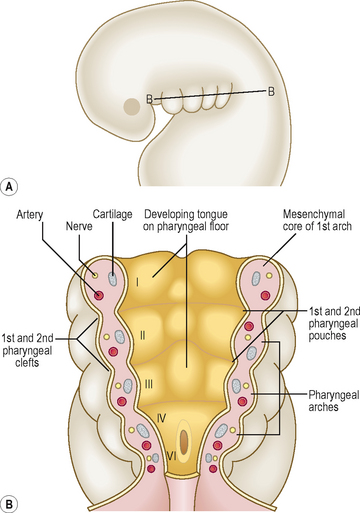
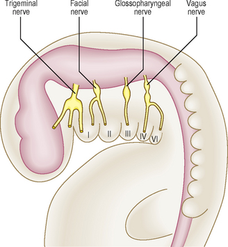
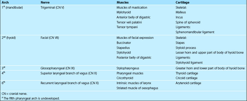
The mesenchyme in each pharyngeal arch differentiates into a bar of cartilage, the associated muscle and an aortic arch artery (Fig. 11.2B). Each pharyngeal arch also contains a cranial nerve (from nerves V, VII, IX and X) that enters it from the brainstem of the developing brain (Fig. 11.3). The cranial nerves carry the motor fibres to supply the muscles derived from the pharyngeal arches, and also carry the sensory fibres to the developing skin covering them and the mucosal tissue lining them. During further development many pharyngeal arch muscles migrate from their original site of origin to reach their final destination, but they retain their original innervation. Thus, the origin of each muscle can be determined from its nerve supply. The adult derivatives of pharyngeal arch structures, including the cartilages, the muscles and their appropriate cranial nerves, are summarized in Table 11.1. The derivatives of pharyngeal arch arteries are described in Chapter 6.
 Clinical box
Pharyngeal clefts
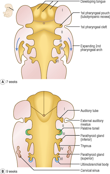
The four pharyngeal clefts separate the pharyngeal arches externally (Fig. 11.4). The first pair of pharyngeal clefts is the only one that contributes to adult structures, namely the external acoustic meatus. The second pharyngeal arch enlarges and grows rapidly as a flap over the remaining three pharyngeal clefts. This flap contains the platysma muscle and fuses below with the epicardial bulge covering the heart. It is possible that remnants of lower clefts lined with ectoderm may remain beneath the flap, forming a cervical sinus, but this is normally obliterated (Fig. 11.4B).
Pharyngeal pouches
The endoderm of the pharyngeal part of the foregut grows laterally as pockets on each side of the pharynx. These paired diverticula, the pharyngeal pouches, develop between the arches (Fig. 11.4A). The first pair of pouches, for example, lies in the interval between the first and second pharyngeal arches. The first four pouches are well developed; the fifth is often absent or rudimentary. The ends of the third and fourth pouches each form a dorsal and a ventral part. The first pouch expands into a tubotympanic recess; the tympanic (middle ear) cavity and auditory tube are derived from this recess (Fig. 11.4). The tympanic membrane is formed by the ectoderm of the first pharyngeal cleft, an intervening layer of mesenchyme and the endoderm lining the pouch. The derivatives of pharyngeal pouches are shown in Table 11.2 and Figure 11.4.
Table 11.2 Main derivatives of the pharyngeal pouches
| Pharyngeal pouch | Main derivative |
|---|---|
| First | Tubotympanic recess, middle ear cavity, auditory tube and tubal tonsil |
| Second | Palatine tonsil (also pharyngeal and lingual tonsils) |
| Third | |
| Fourth | |
| Fifth | Ultimobranchial body: fuses with lateral lobes of the thyroid gland; gives rise to the parafollicular cells (C cells) which produce calcitonin |
 Clinical box
Development of the tongue
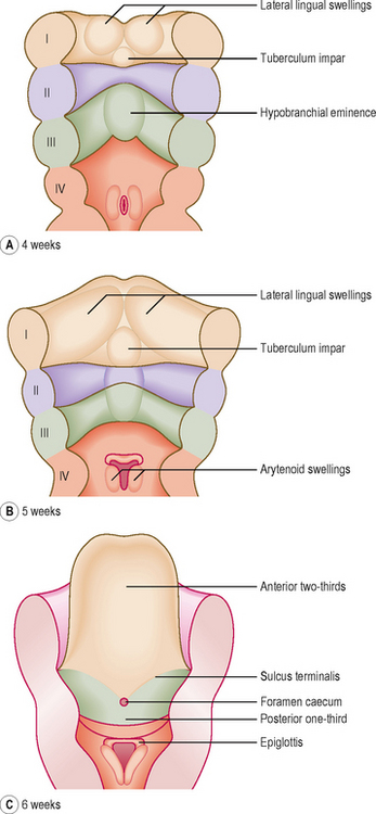
In the fourth week, the tongue develops from mesenchymal swellings covered with ectoderm and endoderm on the floor of the pharynx. The three swellings derived from the first arch mesenchyme, the lateral lingual swellings and a median tuberculum impar, merge with each other to form the anterior two-thirds of the tongue. The second pharyngeal arch makes no contribution to the tongue (Fig. 11.5A, B). Thus, the posterior one-third of the tongue comes from a single swelling, the hypobranchial eminence, derived from the third and fourth pharyngeal arches. A V-shaped groove, the sulcus terminalis, represents the line of fusion between the epithelium covering the first and third pharyngeal arches (Fig. 11.5C). A midline depression at the apex of the sulcus terminalis, the foramen caecum, marks the origin of the thyroid gland (see below).
Stay updated, free articles. Join our Telegram channel

Full access? Get Clinical Tree


