Dedifferentiated Liposarcoma
Thomas Mentzel, MD
Key Facts
Terminology
Malignant lipogenic neoplasm showing abrupt or gradual transition from atypical lipomatous tumor to nonlipogenic sarcoma of variable histology
Clinical Issues
Retroperitoneum, intraabdominal cavity more frequently involved than extremities, spermatic cord, head/neck region, trunk
Recurs locally in at least 40% of cases
Distant metastases observed in 15-20% of cases
Anatomic location represents most important prognostic factor
Dedifferentiation most probably represents time-dependent phenomenon
Dedifferentiation occurs in ˜ 10% of cases of atypical lipomatous tumor
Amount and morphological grade of nonlipogenic areas not of prognostic importance
Microscopic Pathology
Atypical lipogenic tumor cells
Nonlipogenic sarcoma cells
Broad variation of nonlipogenic component
Nonlipogenic component may show low- or high-grade sarcomatous changes
Abrupt or gradual transition from atypical lipomatous tumor (of any subtype) to nonlipogenic sarcoma
Heterologous differentiation in ˜ 10% of cases
Ancillary Tests
Amplification of 12q13-21 region similar to changes in atypical lipomatous tumor
MDM2 and CDK4 overexpression
TERMINOLOGY
Abbreviations
Dedifferentiated liposarcoma (DDLS)
Definitions
Malignant lipogenic neoplasm with abrupt or gradual transition from atypical lipomatous tumor to nonlipogenic sarcoma of variable histology
CLINICAL ISSUES
Epidemiology
Incidence
Occurs in ˜ 10% of cases of atypical lipomatous tumor
90% arise de novo, 10% in local recurrences
Retroperitoneum, intraabdominal cavity are more frequently involved than extremities, spermatic cord, head/neck region, trunk
Mainly in deep soft tissues
Very rare in subcutaneous and dermal tissues
Probably represents time-dependent phenomenon
Age
Middle-aged to elderly patients
Gender
M = F
Presentation
Large, painless mass
Slowly growing neoplasm
Often longstanding mass exhibiting recent increase in size
Treatment
Surgical approaches
Complete excision with tumor-free margins
Prognosis
Recurs locally in ≥ 40% of cases
Distant metastases observed in 15-20% of cases
Overall mortality 25-30% at 5-year follow-up
Anatomic location is most important prognostic factor
Retroperitoneal, intraabdominal lesions exhibit worst clinical behavior
Superficial neoplasms have good prognosis
Amount and morphological grade of nonlipogenic areas not of prognostic importance
IMAGE FINDINGS
General Features
Best diagnostic clue
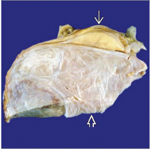
Coexistence of fatty and non-fatty-solid components
Location
Retroperitoneum, intraabdominal cavity, deep soft tissues
Size
Large size, usually > 5 cm
Morphology
Circumscribed
MACROSCOPIC FEATURES
General Features
Large, multinodular yellow mass with solid, often tangray or gray-white nonlipomatous areas
Sections to Be Submitted
Sections of both components must be sampled carefully
Size
May reach large size, especially in abdomen and retroperitoneum
MICROSCOPIC PATHOLOGY
Histologic Features
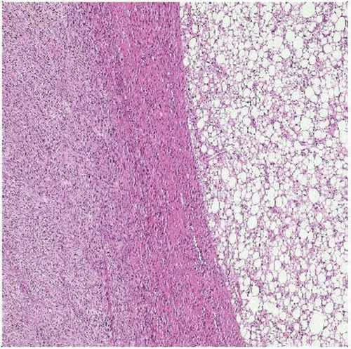
Abrupt or gradual transition from atypical lipomatous tumor (of any subtype) to nonlipogenic sarcoma
Varying size and shape of lipogenic tumor cells
Presence of enlarged and hyperchromatic nuclei in lipogenic component
Nonlipogenic sarcoma component shows broad morphologic variation
High-grade nonlipogenic sarcoma component (pleomorphic sarcoma, intermediate- to high-grade myxofibrosarcoma-like areas)
Often presence of multinucleated giant cells
Increased proliferative activity
Low-grade nonlipogenic component (uniform fibroblastic spindle cells with mild nuclear atypia)
Heterologous differentiation in ˜ 10% of cases
Rhabdomyosarcoma
Leiomyosarcoma
Osteo- or chondrosarcoma
Angiosarcoma
Stay updated, free articles. Join our Telegram channel

Full access? Get Clinical Tree


