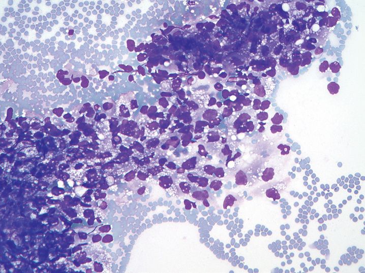
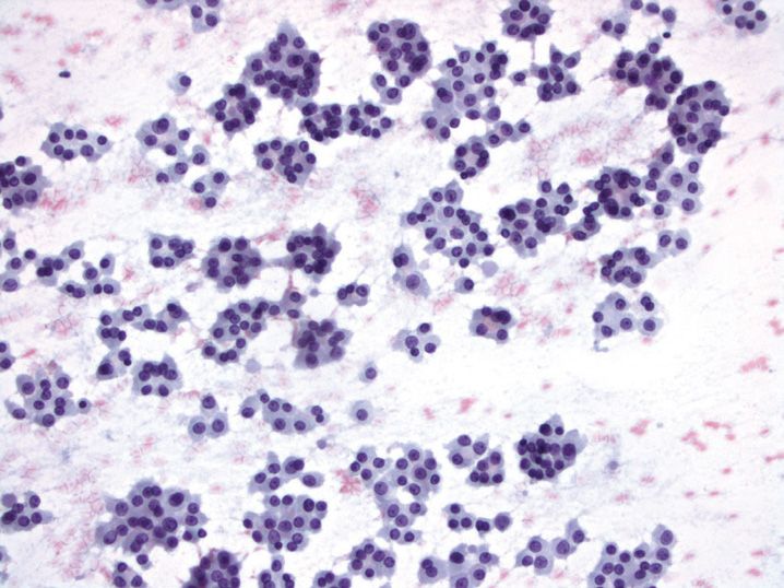
FIGURE 9-1
(A) Alveolar macrophages
(B) Benign bronchial cells
(C) Non-small cell carcinoma
(D) Pleural mesothelial cells
(E) Small cell undifferentiated carcinoma
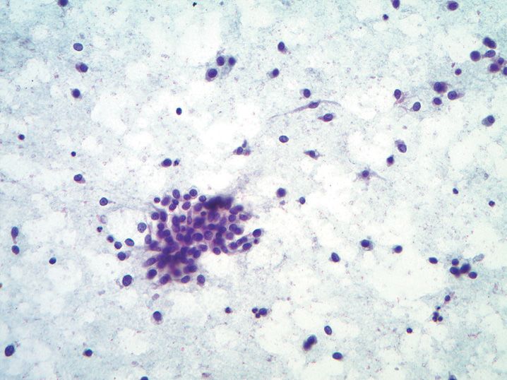
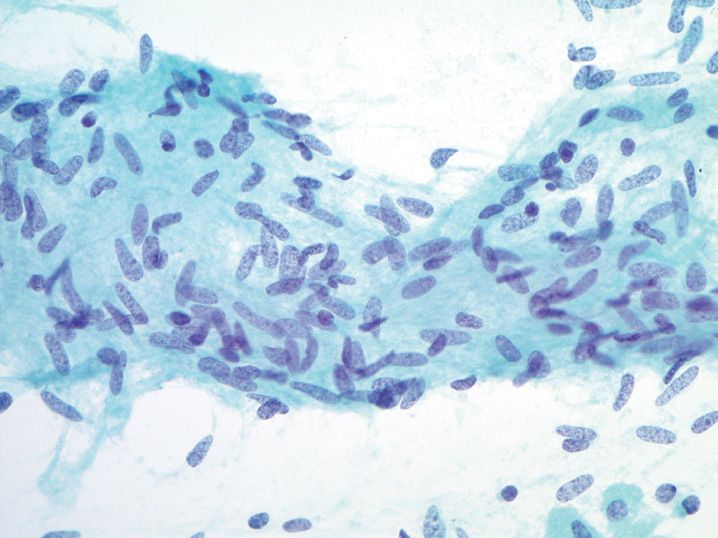
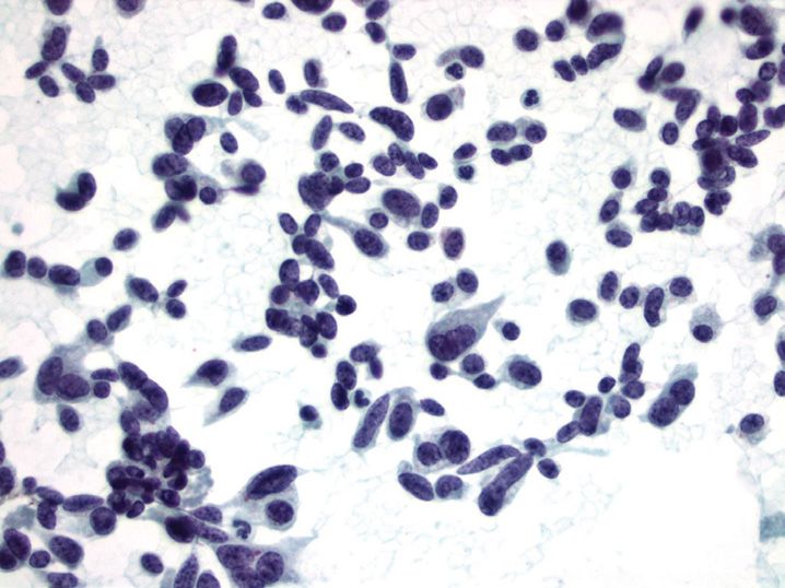
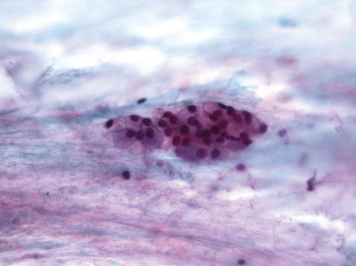
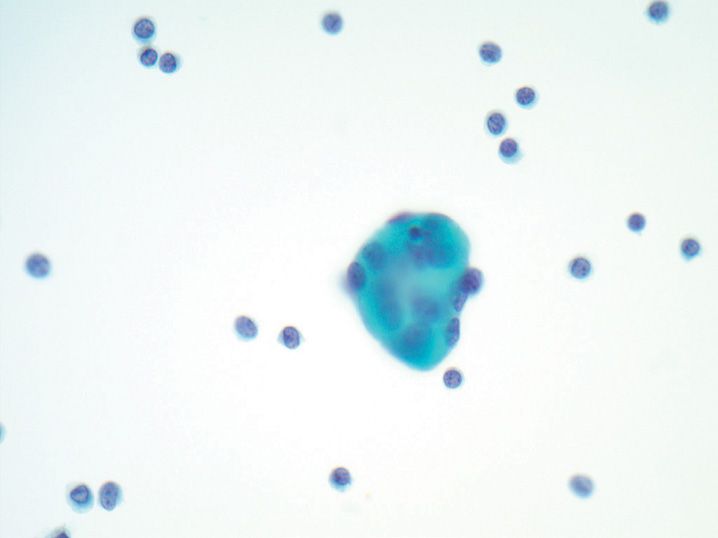
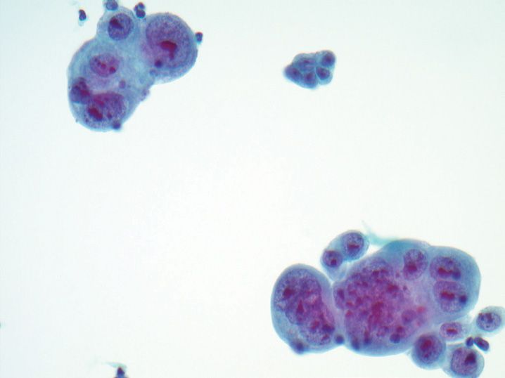
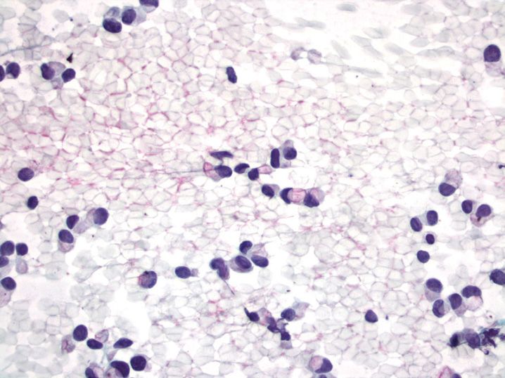
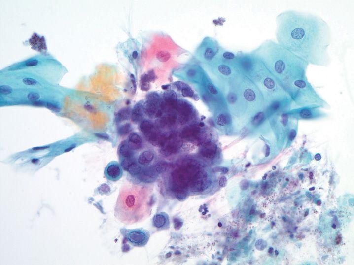
2. A 73-year-old man undergoes transthoracic fine-needle aspiration biopsy of a lung mass (see Figure 9-2). What is the best diagnosis?
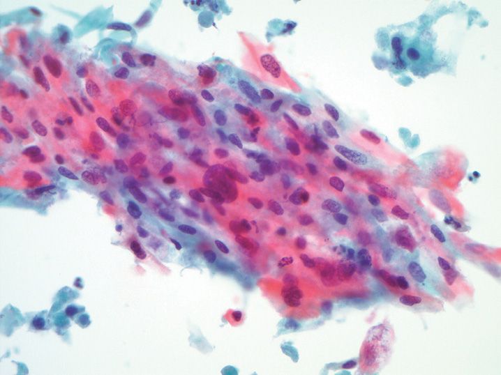
(A) Adenocarcinoma
(B) Reserve cell hyperplasia
(C) Small cell undifferentiated carcinoma
(D) Squamous cell carcinoma
(E) Squamous metaplasia with reactive changes
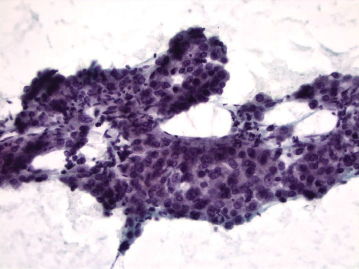
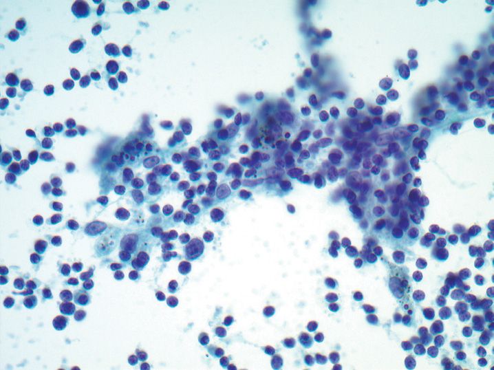
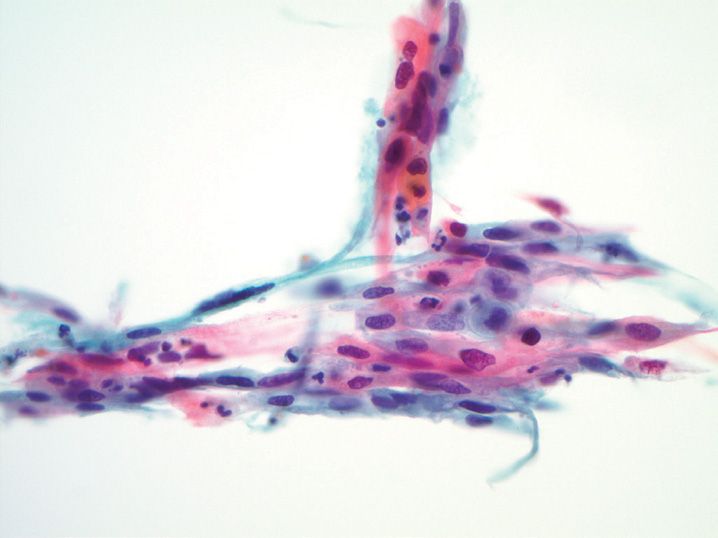
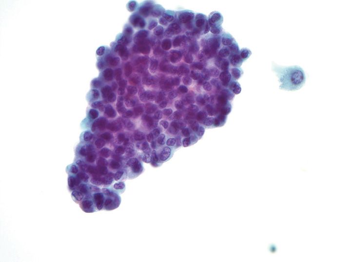
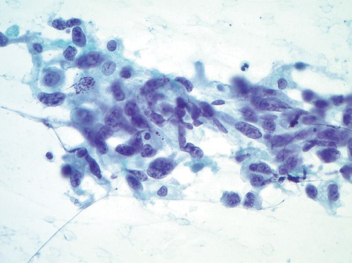
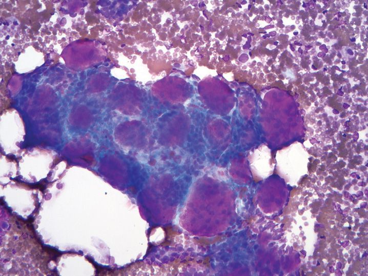
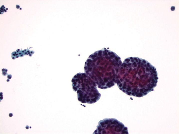
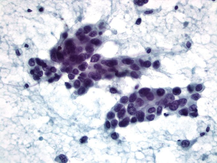
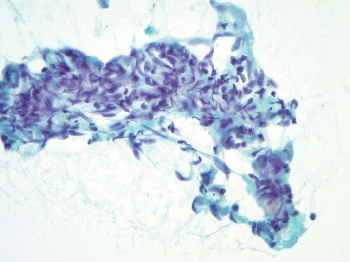
3. A 43-year-old woman presents with an endobronchial mass and undergoes fine-needle aspiration biopsy (see Figure 9-3). What is the best diagnosis for this lesion?
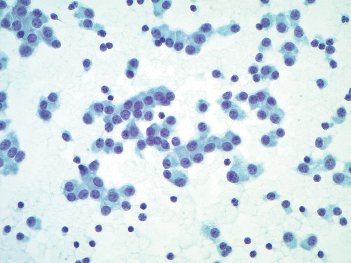
(A) Adenoid cystic carcinoma
(B) Pulmonary carcinoid tumor
(C) Plasmacytoma
(D) Small cell undifferentiated carcinoma
(E) Well-differentiated adenocarcinoma
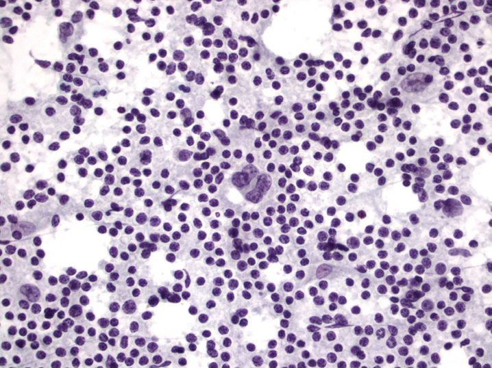
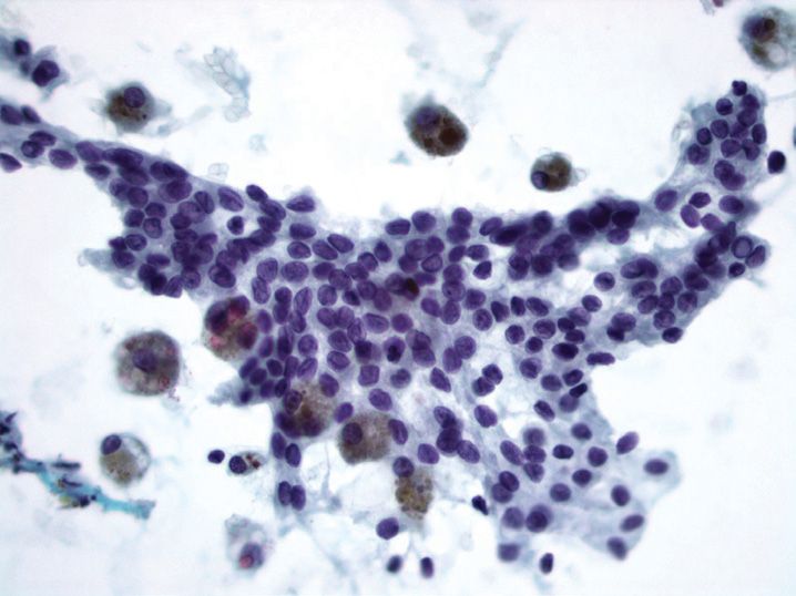
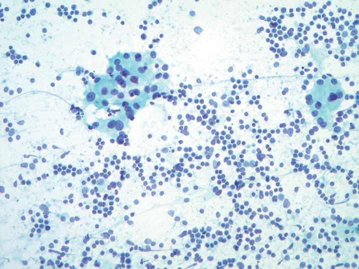
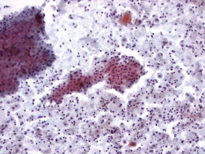
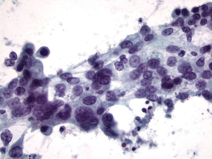
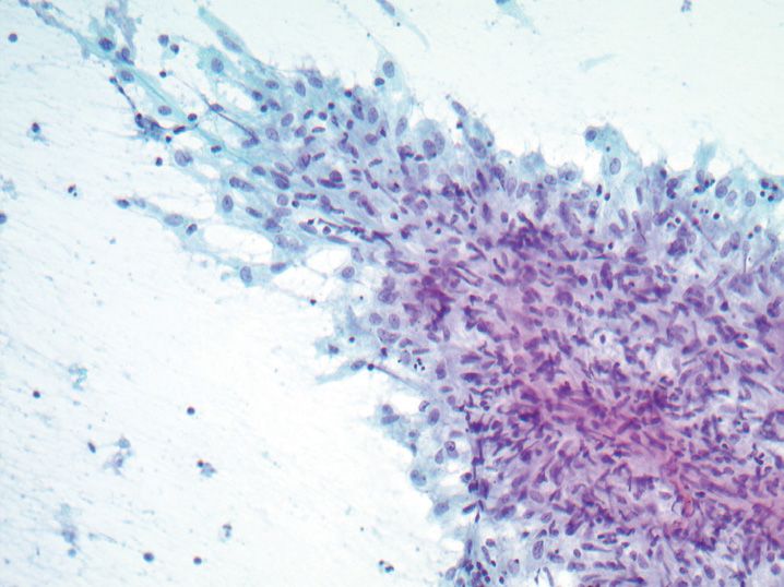
4. A 75-year-old man with a 100-pack-year smoking history presents with a large hilar lung mass. Fine-needle aspiration biopsy is performed (see Figure 9-4). Which of the following immunohistochemical stains, if positive, would confirm the diagnosis?
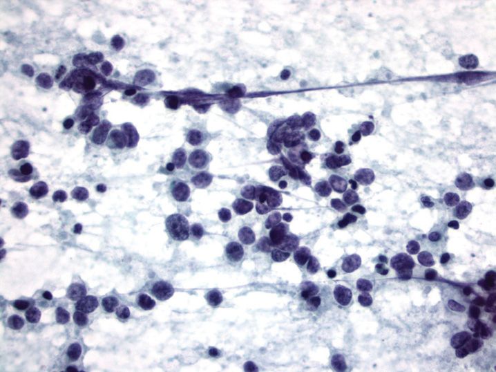
(A) CD20
(B) CD34
(C) Napsin A
(D) P63
(E) Synaptophysin
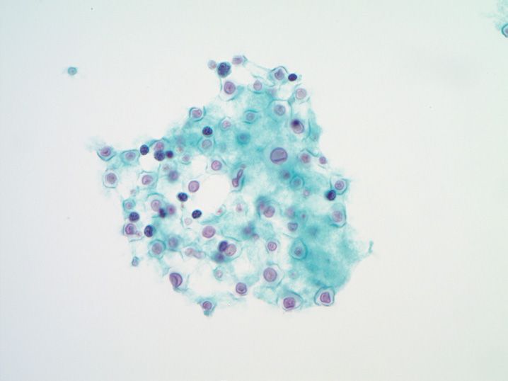
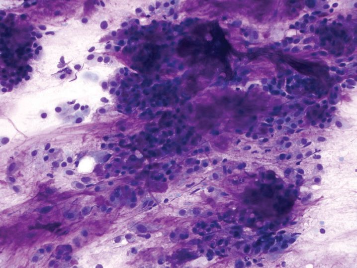
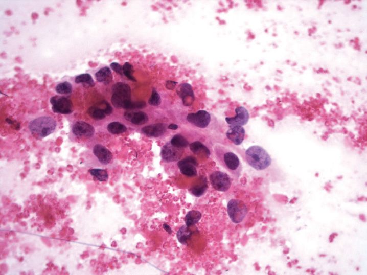
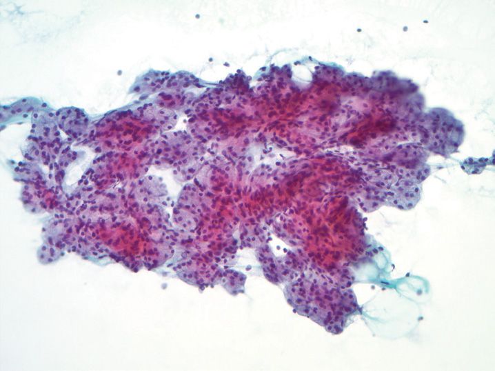
5. A 45-year-old man undergoes fine-needle aspiration biopsy of a renal mass. The following structure is seen in the aspirate sample (see Figure 9-5). From which of the following tissues does this structure likely originate?
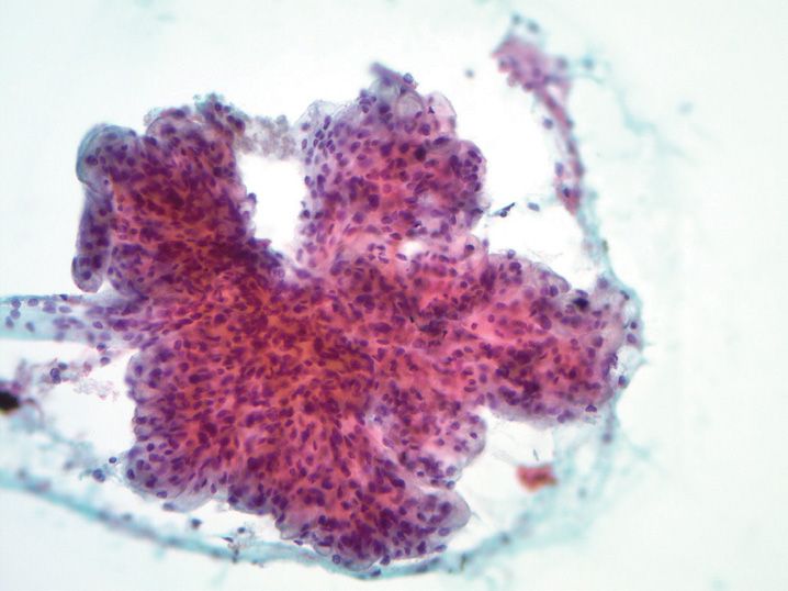
(A) Benign renal cortex
(B) Benign renal medulla
(C) Clear cell renal cell carcinoma
(D) Papillary renal cell carcinoma
(E) Renal medullary carcinoma
6. An 82-year-old man presents with a renal mass.
Fine-needle aspiration biopsy is performed (see Figure 9-6). What cytogenetic abnormality is mostly likely present in this tumor?
(A) Loss of chromosome 3 short arm (3p-)
(B) Loss of chromosome Y
(C) Trisomy 7
(D) TSC1 gene inactivation
(E) Xp11.2 alterations
7. A 72-year-old man with a history of a “kidney tumor” presents with enlarged retroperitoneal lymph nodes. A fine-needle aspiration biopsy is performed (see Figure 9-7). Which of the following tumors is most likely to have this appearance?
(A) Angiomyolipoma
(B) Clear cell renal cell carcinoma
(C) Oncocytoma
(D) Papillary renal cell carcinoma
(E) Urothelial carcinoma
8. A 44-year-old man with a history of cirrhosis is found to have a 3.0-cm liver mass. Fine-needle aspiration biopsy was performed (see Figure 9-8). What is the best diagnosis?
(A) Cholangiocarcinoma
(B) Focal nodular hyperplasia
(C) Metastatic colorectal carcinoma
(D) Regenerating cirrhotic nodule
(E) Well-differentiated hepatocellular carcinoma
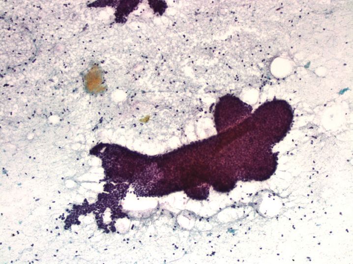
9. A 37-year-old man presents with dry cough, fever, and bilateral pulmonary infiltrates. A bronchioloalveolar lavage is performed (see Figure 9-9, inset Gomori methenamine silver stain). What condition is this process associated with?
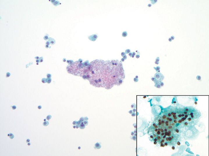
(A) Asbestos exposure
(B) Asthma
(C) Bird handling (e.g., pigeons)
(D) Heavy smoking history
(E) Suppressed immune function
10. A 62-year-old man presents with upper gastrointestinal bleeding. At endoscopy, a polypoid submucosal mass is present in the body of the stomach, with relatively normal overlying mucosa. Endoscopic fine-needle aspiration biopsy is performed (see Figure 9-10). What immunohisto-chemical stain will confirm the diagnosis?
(A) C-kit (CD117)
(B) Cytokeratin
(C) Desmin
(D) HMB-45
(E) Myogenin
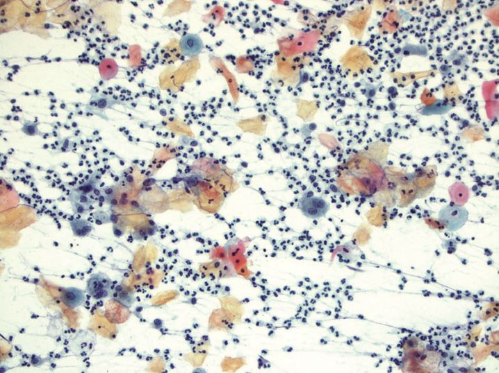
11. A 37-year-old man with poor dentition presents with enlarged right cervical neck lymph nodes. Fine-needle aspiration biopsy is performed (see Figure 9-11). Assuming this figure is representative of the entire aspirate sample, what is the best diagnosis?
(A) Diffuse large B-cell lymphoma
(B) Mantle cell lymphoma
(C) Metastatic squamous cell carcinoma
(D) Reactive lymphoid hyperplasia
(E) Sarcoidosis
12. A 27-year-old man presents with bulky cervical lymphadenopathy, night sweats, and weight loss. Fine-needle aspiration biopsy of a neck lymph node is performed (see Figure 9-12). What is the next best step in the workup of this aspirate sample?
(A) Culture for acid-fast bacteria and fungi
(B) Flow cytometry
(C) Immunohistochemical staining for CD30, CD15, and CD20
(D) Immunohistochemical staining for cytokeratin and S-100
(E) Serologic studies for Epstein–Barr virus
13. A 45-year-old man presents with headache and neck stiffness. A cerebrospinal fluid sample is submitted for cytologic examination (see Figure 9-13). What special stain will highlight the capsule of this organism?
(A) Fontana–Masson
(B) Gomori methenamine silver (GMS)
(C) Mucicarmine
(D) Periodic acid Schiff (PAS)
(E) Ziehl–Neelson
14. A 44-year-old man undergoes fine-needle aspiration biopsy (FNA) of a solitary 2.5-cm solid thyroid nodule (see Figure 9-14). Assuming this figure is representative of the entire aspirate, what is the most appropriate next step in management of this lesion?
(A) Clinical follow-up only with ultrasound in 1 year
(B) Repeat FNA in 3 months
(C) Repeat FNA within a week
(D) Thyroid lobectomy
(E) Total thyroidectomy with central neck lymph node dissection
15. A 43-year-old man who recently emigrated from Egypt has a bladder mass, and a urine sample is submitted for cytologic examination (see Figure 9-15). This neoplasm is associated with what infectious organism?
(A) Clonorchis sinensis
(B) Human papilloma virus
(C) Polyomavirus
(D) Schistosoma hematobium
(E) Schistosoma mansoni
16. A 71-year-old woman presents with hematuria. Cystoscopy of the bladder is negative for abnormalities, and a urine specimen at the time of cystoscopy is submitted for cytologic examination (see Figure 9-16). What is the most likely diagnosis?
(A) Flat urothelial carcinoma in situ
(B) High-grade papillary urothelial carcinoma of the bladder
(C) Nephrolithiasis
(D) Polyoma virus infection
(E) Squamous cell carcinoma of the bladder
17. A 29-year-old woman undergoes fine-needle aspiration biopsy for a 1.8-cm thyroid nodule (see Figure 9-17). The most important cytologic feature for identification and diagnosis of this lesion is
(A) Absence of colloid
(B) Abundant cytoplasm
(C) Architectural arrangement of follicular cells
(D) Background of cyst contents and multinucleated giant cells
(E) Nuclear atypia
18. A 33-year-old man undergoes fine-needle aspiration biopsy of a 1.0-cm right upper pole thyroid nodule (see Figure 9-18). His brother was recently diagnosed with the same neoplasm. What is the most likely mutation present in this lesion?
(A) BRAF point mutation
(B) PAX8/PPARγ translocation
(C) RAS point mutation
(D) RET point mutation
(E) RET/PTC translocation
19. An 89-year-old man presents with a rapidly growing thyroid mass causing respiratory stridor. The mass is very firm to palpation. A fine-needle aspiration biopsy is performed (see Figure 9-19). What is the most likely diagnosis?
(A) Benign, reactive changes
(B) Metastatic squamous cell carcinoma
(C) Papillary thyroid carcinoma
(D) Pleomorphic sarcoma of the thyroid gland
(E) Undifferentiated (anaplastic) thyroid carcinoma
20. A 41-year-old woman undergoes thyroid fine-needle aspiration biopsy of a thyroid gland nodule (see Figure 9-20). What peripheral blood test is likely to be abnormal in this patient?
(A) Elevated serum calcitonin
(B) Elevated serum calcium
(C) Elevated serum thyroglobulin
(D) Elevated T3/T4 levels
(E) Positive antithyroglobulin antibodies
21. A 62-year-old man presents with a parotid mass. Fine-needle aspiration biopsy is performed (see Figure 9-21). What is the best diagnosis for this lesion?
(A) Acinic cell carcinoma
(B) Adenoid cystic carcinoma
(C) Mucoepidermoid carcinoma
(D) Pleomorphic adenoma
(E) Warthin tumor
22. A 14-year-old boy presents with a parotid mass, and undergoes fine-needle aspiration biopsy (see Figure 9-22). All of the following statements are true regarding this lesion except
(A) Associated with a recurring translocation of chromosomes 11 and 19
(B) Can arise primarily within the mandibular bone
(C) Frequently a cause of false-negative aspirate results
(D) May appear oncocytic in fine-needle aspiration biopsy samples
(E) Second most common malignant salivary gland neoplasm in children
23. A 55-year-old woman presents with a submandibular mass, and undergoes fine-needle aspiration biopsy (see Figure 9-23). What is the most likely diagnosis?
(A) Acinic cell carcinoma
(B) Adenoid cystic carcinoma
(C) Mucoepidermoid carcinoma
(D) Pleomorphic adenoma
(E) Warthin tumor
24. A 58-year-old man has bilateral parotid lesions, and undergoes fine-needle aspiration biopsy (see Figure 9-24). This lesion is highly associated with what risk factor?
(A) Alcohol
(B) Human papilloma virus
(C) Radiation exposure
(D) Sjögren syndrome
(E) Smoking
25. A 76-year-old man undergoes fine-needle aspiration biopsy of a parotid mass (see Figure 9-25). Immunohistochemical staining on the cell block demonstrates 3+ continuous membranous staining for HER2. What is the most likely diagnosis?
(A) Adenocarcinoma, favor breast origin
(B) Anaplastic large cell lymphoma
(C) High-grade mucoepidermoid carcinoma
(D) Malignant melanoma
(E) Salivary duct carcinoma
26. A 52-year-old woman with an ovarian mass undergoes salpingo-oophorectomy. At the time of surgery, a pelvic washing is performed, which shows the following finding (see Figure 9-26). What is the best diagnosis for this washing?
(A) Atypical cells, suspicious for a borderline serous tumor
(B) Atypical cells, suspicious for malignant mesothelioma
(C) Negative for malignancy
(D) Positive for malignancy, malignant mesothelioma
(E) Positive for malignancy, papillary serous carcinoma
27. A 65-year-old woman presents with a large right pleural effusion. Cytologic examination of the fluid demonstrates the following changes (see Figure 9-27). What is the most likely origin of these cells?
(A) Ductal carcinoma of the breast
(B) Gastric carcinoma
(C) Lobular carcinoma of the breast
(D) Lung carcinoma
(E) Reactive mesothelial cells
28. A 72-year-old woman presents with obstructive jaundice, and imaging shows a solid pancreatic head mass with irregular contours. Fine-needle aspiration biopsy is performed (see Figure 9-28). What is the best diagnosis for this aspirate?
(A) Acinar cell carcinoma
(B) Chronic pancreatitis
(C) Ductal adenocarcinoma
(D) Intraductal papillary mucinous neoplasm
(E) Pancreatic endocrine neoplasm
29. A 61-year-old woman undergoes endoscopic ultrasound-guided fine-needle aspiration biopsy of a pancreatic tail mass (see Figure 9-29). What is the most likely diagnosis based on this cytomorphologic appearance?
(A) Acinar cell carcinoma
(B) Chronic pancreatitis
(C) Ductal adenocarcinoma
(D) Intraductal papillary mucinous neoplasm
(E) Pancreatic endocrine neoplasm
30. A 68-year-old woman undergoes total hysterectomy and salpingo-oophorectomy for an ovarian mass. Pathologic examination demonstrates a high-grade papillary serous carcinoma confined to one ovary. A pelvic washing performed at the time of surgery shows the following change (see Figure 9-30). What effect does the pelvic washing finding have on the stage of her ovarian cancer?
(A) No change to stage
(B) Upstage from T1a to T1c
(C) Upstage from T1 to T3
(D) Upstage from N0 to N1
(E) Upstage from M0 to M1
31. A 37-year-old woman undergoes fine-needle aspiration biopsy of a breast lesion (see Figure 9-31). Based on the cytologic features present, what is the most likely diagnosis?
(A) Benign cyst
(B) Ductal carcinoma
(C) Fat necrosis
(D) Fibroadenoma
(E) Intraductal papilloma
32. A 42-year-old woman who is a smoker develops a tender lump underneath the nipple. Fine-needle aspiration biopsy is performed (see Figure 9-32). What is the most likely diagnosis?
(A) Ductal carcinoma
(B) Fibroadenoma
(C) Intraductal papilloma
(D) Metastatic squamous cell carcinoma
(E) Subareolar abscess
33. A 72-year-old woman undergoes fine-needle aspiration biopsy of a breast mass (see Figure 9-33). The best diagnosis for this aspirate sample is
(B) Fibroadenoma
(C) Intraductal papilloma
(D) Lobular carcinoma
(E) Subareolar abscess
34. A 59-year-old woman presents with a breast mass, and undergoes fine-needle aspiration biopsy (see Figure 9-34). What is the best diagnosis for this lesion?
(A) Ductal carcinoma
(B) Fat necrosis
(C) Fibroadenoma
(D) Lobular carcinoma
(E) Metastatic signet ring carcinoma to the breast
35. A 23-year-old man presents with a rapidly growing mass in the lateral neck that measures 1.8 cm and is subcutaneous. Fine-needle aspiration biopsy is performed and is suggestive of nodular fasciitis (see Figure 9-35). All of the following are cytologic features of this lesion except
(A) Acute inflammation
(B) Frequent mitotic figures
(C) Marked nuclear hyperchromasia and atypia
(D) Myxoid background
(E) Spindled or stellate-shaped cells
36. Transbronchial fine-needle aspiration biopsy of a mediastinal lymph node demonstrates the following change (see Figure 9-36). What is the next best step in the evaluation of this lymph node?
(A) Flow cytometry
(B) Gram stain on aspirate smears
(C) Immunohistochemistry on cell block material
(D) Microbiologic culture for fungi and acid-fast bacteria
(E) Serum angiotensin-converting enzyme (ACE) levels
37. A 7-year-old boy from Africa presents with a rapidly enlarging neck mass, and fine-needle aspiration biopsy demonstrates a monotonous population of atypical lymphocytes with dark blue cytoplasm and multiple cytoplasmic vacuoles. What gene is most likely altered in this neoplasm?
(A) BCL2
(B) C-myc
(C) Immunoglobulin light chain
(D) N-myc
(E) T-cell receptor gamma chain
38. All of the following are cytologic features of mesothelial cells in effusion cytology except
(A) Binucleation and multinucleation common
(B) Clusters with cytoplasmic windows between cells
(C) Clusters with smooth community borders
(D) Dense inner cytoplasm with outer pale rim, so-called “lacy skirt”
(E) Round-to-oval nucleus with small nucleolus
39. A 55-year-old woman has a history of invasive ductal carcinoma of the breast, and presents with a large pleural effusion. Atypical cells are present in her pleural effusion. Which of the following panels of immunohistochemical stains would be most useful in discriminating metastatic adenocarcinoma from reactive mesothelial cells in her effusion?
(A) Calretinin, cytokeratins 5/6 (CK5/6), carcinoembryonic antigen (CEA), B72.3
(B) Calretinin, CK5/6, WT-1, D2-40
(C) CEA, B72.3, MOC-31, cytokeratin 7 (CK7)
(D) CK7, AE1/3, CAM5.2
(E) CK7, estrogen receptor, progesterone receptor
40. A 54-year-old man presents with bilateral pleural effusions, cavitary pulmonary lesions, and chronic cough. Examination of his pleural fluid demonstrates nearly 100% small mature lymphocytes and almost no mesothelial cells. What process is the most likely cause of this clinical picture?
(A) Aspergillus fumigatus
(B) Metastatic small cell undifferentiated carcinoma
(C) Mycobacterium tuberculosis
(D) Primary effusion lymphoma
(E) Systemic lupus erythematosus
41. A 28-year-old man presents with a pleural effusion. Examination of the effusion demonstrates numerous eosinophils. Which of the following is a possible cause of this finding?
(A) Empyema
(B) Primary effusion lymphoma
(C) Rheumatoid pleuritis
(D) Spontaneous pneumothorax
(E) Tuberculous pleuritis
42. When examining a pleural or peritoneal effusion cytology sample, all of the following findings are concerning for malignancy except
(A) Large cell clusters (>50 cells)
(B) Mitotic figures
(C) Signet ring vacuoles
(D) Smooth community borders to cell groups
(E) Two distinct cell populations
43. In effusion cytology, some metastatic tumors will consist of predominantly single tumor cells rather than forming clusters. This pattern is called the “dispersed” or “mesothelial-type” pattern of metastasis. All of the following tumors commonly involve effusions with a dispersed pattern except
(A) Lobular breast carcinoma
(B) Malignant melanoma
(C) Non-Hodgkin lymphoma
(D) Ovarian serous carcinoma
(E) Signet ring gastric carcinoma
44. A 32-year-old woman notices a well-circumscribed mass in her breast. Fine-needle aspiration biopsy is performed and shows a cellular aspirate composed of loosely cohesive epithelial cells with foamy cytoplasm and prominent nucleoli, in a background of proteinaceous material and stripped nuclei. These changes are most consistent with what diagnosis?
(A) Ductal carcinoma
(B) Fibroadenoma
(C) Lactating adenoma
(D) Lobular carcinoma
(E) Mucinous carcinoma
45. All of the following are features of fat necrosis on fine-needle aspiration cytology except
(A) Adipocytes
(B) Amorphous granular background debris
(C) Foamy histiocytes
(D) Hypercellular epithelial fragments
(E) Inflammation
46. Which of the following features favors a diagnosis of phyllodes tumor over fibroadenoma on fine-needle aspiration biopsy of the breast?
(A) Branching fragments of ductal epithelium
(B) Hypercellular stromal fragments
(D) Overall high cellularity aspirate sample
(E) Spindle cells with short, oval nuclei
47. A woman undergoes fine-needle aspiration biopsy of a breast lesion, and the aspirate smears demonstrate abundant extracellular mucinous/myxoid material with admixed epithelial cells. All of the following lesions are in the differential diagnosis of this process except
(A) Colloid carcinoma
(B) Ductal carcinoma with mucinous features
(C) Fibroadenoma
(D) Mucocele-like lesion
(E) Tubular carcinoma
48. All of the following are cytologic features of pilomatricoma except
(A) Background of amorphous debris and calcification
(B) Background of naked nuclei
(C) Clusters of basaloid cells with high nuclear-to-cytoplasmic ratio
(D) Coarse hyperchromatic and irregular chromatin
(E) Ghost cells (anucleated squamous cells)
49. All of the following childhood tumors frequently have a small round blue cell appearance on fine-needle aspiration biopsy except
(A) Acute lymphoblastic lymphoma
(B) Ewing sarcoma/primitive neuroectodermal tumor
(C) Infantile fibrosarcoma
(D) Neuroblastoma
(E) Rhabdomyosarcoma
50. Which of the following features is seen in fine-needle aspiration samples of schwannoma?
(A) Epithelioid cells with plasmacytoid cytoplasm and binucleation
(B) Marked nuclear pleomorphism and tumor giant cells with prominent nucleoli
(C) Spindle cells with rounded, cigar-shaped nuclei
(D) Stellate, fibroblast-like spindle cells in an inflammatory background
(E) Wavy spindle cells with tapered nuclei
51. A 45-year-old woman undergoes fine-needle aspiration biopsy of a palpable breast mass. The aspirate smears demonstrate numerous naked nuclei in a granular background, and a few groups of intact polygonal cells have abundant granular cytoplasm and small round nuclei with no significant pleomorphism. What immunohistochemical stain will be positive in this neoplasm?
(A) CD68
(B) Cytokeratin
(C) Desmin
(D) Myogenin
(E) S-100
52. A 49-year-old man presents with a deep thigh mass. A fine-needle aspiration biopsy is performed, and shows a prominent myxoid background with delicate branching vessels, cells with multiple cytoplasmic vacuoles, and round-to-spindled cells with scant cytoplasm. On the basis of this cytologic appearance, what cytogenetic translocation is most likely to be present in this lesion?
(A) t(X;17)(p11;q25)
(B) t(X;18)(p11;q11)
(C) t(2;13)(q35;q14)
(D) t(11;22)(q24;q12)
(E) t(12;16)(q13;p11)
53. Basaloid salivary gland neoplasms can show considerable overlap on fine-needle aspiration biopsy. Which of the following features, if present, would favor a diagnosis of basal cell adenoma?
(A) Fibrillary metachromatic matrix material
(B) High mitotic activity and necrosis
(C) Spheres of acellular, sharply demarcated meta-chromatic matrix material
(D) Squamous differentiation with nuclear atypia
(E) Thick ribbon of matrix material surrounding cell groups with peripheral palisading
54. Fine-needle aspiration biopsy of a parotid mass demonstrates polygonal cells with abundant cytoplasm and round central nucleoli. All of the following salivary gland lesions are in the differential diagnosis except
(A) Acinic cell carcinoma
(B) Adenoid cystic carcinoma
(C) Mucoepidermoid carcinoma
(D) Oncocytoma
(E) Warthin tumor
55. A parotid mass fine-needle aspiration biopsy contains extracellular metachromatic matrix material on Diff-Quik stain. Which of the following tumors can have this appearance on aspiration biopsy?
(A) Acinic cell carcinoma
(B) Epithelial myoepithelial carcinoma
(C) Mucoepidermoid carcinoma
(D) Myoepithelioma
(E) Salivary duct carcinoma
56. Which of the following salivary gland tumors is least likely to appear cystic on imaging?
(A) Acinic cell carcinoma
(B) Lymphoepithelial cyst
(C) Polymorphous low-grade adenocarcinoma
(D) Mucoepidermoid carcinoma
(E) Warthin tumor
57. According to the 2007 Bethesda System for Reporting Thyroid Cytopathology, all of the following thyroid fine-needle aspirates would be considered adequate for interpretation except
(A) Abundant colloid only
(B) Abundant cyst contents only
(C) Changes of chronic lymphocytic thyroiditis with 5 groups of 10 follicular cells
(D) Only 3 groups of follicular cells with nuclear grooves, fine chromatin, and a nuclear pseudoinclusion
(E) Scant colloid and 8 groups of 10 benign follicular cells
58. An interpretation of “suspicious for follicular neoplasm, Hürthle cell type” on a thyroid fine-needle aspiration biopsy requires all of the following cytologic findings except
(A) Absence of lymphocytes
(B) Absence of nuclear features of papillary thyroid carcinoma
(C) At least 50% of the follicular cells have Hürthle cell change
(D) Follicular cells with abundant cytoplasm, large nuclei, and prominent nucleoli
(E) Minimal colloid
59. A 32-year-old woman undergoes fine-needle aspiration biopsy of a solitary thyroid nodule. The cytopathologist makes an interpretation of “atypia of undetermined significance.” According to the 2007 Bethesda System for Reporting Thyroid Cyto-pathology recommendations, at excision what percentage of patients with this interpretation will have a malignant neoplasm?
(A) <3%
(B) 5–15%
(C) 15–30%
(D) 60–75%
(E) 97–99%
60. In what clinical setting is a thyroid fine-needle aspiration biopsy considered clinically adequate with an interpretation of “nondiagnostic, cyst fluid only”?
(A) Multilocular cyst 1.5 cm in size
(B) Multilocular cyst 1.0 cm in size with microcalcifications
(C) Solid nodule 2.0 cm in size
(D) Unilocular cyst 2.5 cm in size
(E) Unilocular cyst 4.5 cm in size
61. A 46-year-old woman undergoes fine-needle aspiration biopsy of a 1.5-cm unilocular cyst of the right thyroid. Approximately 5 mL of crystal clear fluid is aspirated, and the cyst collapses. Review of the aspirate smears demonstrates nearly acellular cyst fluid. What ancillary test can the pathologist perform to confirm the diagnosis?
(A) Aspirate fluid calcitonin level
(B) Aspirate fluid parathyroid hormone level
(C) Aspirate fluid thyroglobulin level
(D) Serum calcitonin level
(E) Serum thyroglobulin level
62. A 46-year-old woman submits a urine specimen for cytologic examination. Several large papillary clusters of urothelial cells with a low nuclear-to-cytoplasmic ratio and minimal atypia are present. The differential diagnosis of this process includes all of the following except
(A) Bladder papilloma
(B) High-grade papillary urothelial carcinoma
(C) Low-grade papillary urothelial carcinoma
(D) Nephrolithiasis
(E) Recent instrumentation of the bladder
63. Which of the following features, if present in a urine cytology sample, would favor a diagnosis of polyoma virus infection over a high-grade urothelial carcinoma?
(A) Clusters of atypical cells
(B) Extensive cellular degeneration
(C) High nuclear-to-cytoplasmic ratio
(D) Nuclear hyperchromasia
(E) Round nuclei with smooth contours
64. A 55-year-old man with a history of high-grade urothelial carcinoma is seen for annual follow-up and a urine cytology specimen is submitted for a fluorescence in situ hybridization test (e.g., UroVysion™) to assess for recurrent disease. What type of genetic changes does this assay detect?
(A) Aneuploidy of multiple chromosomes (3, 7, 17)
(B) Break-apart probe for translocations involving chromosome 9p
(C) Fusion signal probe for translocations involving chromosome 17
(D) Homozygous deletion of chromosome 7
(E) Point mutation of chromosome 3
65. A 32-year-old woman presents with headaches, and a cerebrospinal fluid (CSF) sample is submitted for cytologic examination. The CSF sample demonstrates increased numbers of lymphocytes with occasional atypical lymphoid cells. All of the following processes can cause this appearance except
(A) Acute lymphoblastic leukemia with leptomeningeal involvement
(B) Borrelia burgdorferi (Lyme disease) infection
(C) Neisseria meningitidis infection
(D) Primary central nervous system lymphoma
(E) Viral meningitis
66. A premature baby girl with hydrocephalus and seizures has cerebrospinal fluid sample submitted for cytologic examination. Imaging does not demonstrate a mass lesion. The fluid demonstrates clusters of small dark cells with scant cytoplasm and nuclear molding. What is the most likely source of these cells?
(A) Germinal matrix cells
(B) Medulloblastoma
(C) Neuroblastoma
(D) Primary CNS lymphoma
(E) Small cell carcinoma
67. A sputum sample is submitted for cytologic examination in the workup of a patient with a lung mass. What cellular component is required to consider the sputum sample adequate?
(A) Alveolar macrophages
(B) Ciliated bronchial cells
(C) Goblet cells
(D) Mature squamous cells
(E) Metaplastic squamous cells
68. In a bile duct brushing, which of the following features would favor a diagnosis of adenocarcinoma over reactive changes?
(A) Abundant cytoplasm
(B) Cohesive clusters
(C) Large nuclei
(D) Marked nuclear size variation
(E) Prominent nucleoli
69. A woman presents with nipple discharge. Which of the following nipple discharge characteristics is most associated with an underlying malignancy?
(A) Bilateral discharge
(B) Bloody discharge
(C) Milky discharge
(D) Postpartum discharge
(E) Purulent discharge
70. A 45-year-old woman presents with unilateral bloody nipple discharge. A nipple discharge smear is made, and shows multiple cohesive three-dimensional clusters of small ductal cells with mild nuclear pleomorphism and scant cytoplasm. What is the best diagnosis for this cytology sample?
(A) Fibroadenoma
(B) Negative for malignancy, fibrocystic change
(C) Papillary lesion
(D) Positive for malignancy, ductal carcinoma
(E) Positive for malignancy, lobular neoplasia
71. Patients with Barrett’s esophagus are at increased risk of developing invasive adenocarcinoma. What is the characteristic feature of Barrett’s esophagus on esophageal brushing cytology?
(A) Atypical squamous cells
(B) Benign squamous cells
(C) Goblet cells
(D) Necrosis
(E) Sheets of columnar glandular epithelium
72. In a patient with known Barrett’s esophagus, which of the following features would favor a diagnosis of high-grade dysplasia or adenocarcinoma over repair on an esophageal brushing cytology?
(A) Columnar glandular cells with interspersed goblet cells
(B) Honeycomb appearance
(C) Mitotic figures
(D) Numerous single atypical cells
(E) Vesicular chromatin with prominent nucleoli
73. Molecular testing for which of the following gene alterations can be performed on cytologic samples of metastatic colorectal carcinoma to determine tumor sensitivity to antiepidermal growth factor receptor (anti-EGFR) therapy?
(A) Epidermal growth factor receptor (EGFR) gene amplification
(B) K-ras point mutation
(C) P53 gene amplification
(D) RET point mutation
(E) Vascular endothelial growth factor (VEGF) point mutation
74. An 18-year-old woman presents with malaise, pharyngitis, and bilateral cervical neck lymphadenopathy. Serologic studies demonstrate elevated IgM titers for Epstein–Barr virus. If fine-needle aspiration biopsy of a neck lymph node is performed, what is the most likely pattern seen?
(A) Epithelioid histiocytes and well-formed granulomas
(B) Extensive necrosis and marked acute inflammation
(C) Monotonous large atypical lymphocytes with prominent nucleoli
(D) Monotonous small mature lymphocytes lacking tingible body macrophages
(E) Polymorphous lymphoid population containing abundant plasma cells and plasmacytoid lymphocytes
75. A man with a history of malignant melanoma undergoes fine-needle aspiration biopsy of an enlarged lymph node. All of the following are cytomorphologic features of malignant melanoma except
(A) Binucleation with “mirror image” nuclei
(B) Cohesive cell clusters
(C) Large nuclei with prominent macronucleoli
(D) Nuclear pseudoinclusions
(E) Plasmacytoid appearance (eccentric nuclei)
76. All of the following are features of anaplastic large cell lymphoma on fine-needle aspiration biopsy except
(A) Horseshoe-shaped nuclei with eccentric cytoplasm
(B) Immunohistochemical staining for CD30
(C) Large nuclei with prominent nucleoli
(D) Marked nuclear pleomorphism
(E) Background containing numerous lymphoglandular bodies
77. Merkel cell carcinoma is difficult to distinguish from small cell undifferentiated carcinoma of the lung on fine-needle aspiration biopsy. What immunohistochemical stain will be positive in Merkel cell carcinoma, but negative in small cell undifferentiated carcinoma of the lung?
(A) CD56
(B) Chromogranin
(C) Cytokeratin 20
(D) Synaptophysin
(E) Thyroid transcription factor 1
78. Fine-needle aspiration biopsy of a liver mass demonstrates abundant normal-appearing hepatocytes arranged in small clusters and as single cells, without endothelial wrapping or transgressing vessels. No bile duct epithelium is seen. All of the following are possible correlates to this appearance except
(A) Focal nodular hyperplasia
(B) Hepatic adenoma
(C) Hepatocellular carcinoma, fibrolamellar variant
(D) Normal liver, the lesion was missed
(E) Regenerating nodule of cirrhosis
79. Which of the following features, if seen on a liver fine-needle aspiration biopsy sample, would favor a diagnosis of well-differentiated hepatocellular carcinoma over a regenerating cirrhotic nodule?
(A) Bile pigment
(B) Marked variation in nuclear size
(C) Nuclear pseudoinclusions
(D) Prominent nucleoli
(E) Thickened trabeculae
80. All of the following are cytomorphologic features of metastatic colorectal carcinoma to the liver on fine-needle aspiration biopsy except
(A) Dirty necrosis
(B) Hyperchromatic chromatin
(C) Low cuboidal cells
(D) Oval-to-cigar-shaped nuclei
(E) Palisaded or picket-fence nuclear arrangement
81. A 45-year-old woman presents with a cystic pancreatic lesion. Which of the following features would favor a diagnosis of intraductal papillary mucinous neoplasm over a mucinous cystic neoplasm?
(A) Atypical mucinous epithelium
(B) Background of extracellular mucin
(C) Connection with the main pancreatic duct
(D) Elevated carcinoembryonic antigen (CEA) level in the cyst fluid
(E) Location in the pancreas body
82. An endoscopic ultrasound-guided fine-needle aspiration biopsy of a pancreatic lesion is performed. Which of the following features would favor a diagnosis of well-differentiated pancreatic ductal adenocarcinoma over reactive changes in chronic pancreatitis?
(A) Anisonucleocytosis
(B) Cohesive epithelial sheets
(C) Maintained nuclear polarity
(D) Prominent nucleoli
(E) Scattered mitotic figures
83. Which of the following techniques is the most sensitive, specific, and cost-effective for the evaluation and diagnosis of pancreatic lesions?
(A) Computed tomography (CT)-guided fine-needle aspiration biopsy
(B) Diagnostic surgical excision
(C) Endoscopic ultrasound guided fine-needle aspiration biopsy (EUS-FNA)
(D) Laparoscopic biopsy
(E) Transabdominal ultrasound-guided fine-needle aspiration biopsy
84. Fine-needle aspiration biopsy of a 3.0-cm pancreatic cyst yields macrophages only. Ancillary testing with carcinoembryonic antigen (CEA) and amylase is performed on 2 mL of cyst fluid. Which of the following laboratory results would favor a diagnosis of pseudocyst?
(A) CEA 5 ng/mL, amylase high
(B) CEA 4 ng/mL, amylase low
(C) CEA 220 ng/mL, amylase high
(D) CEA 500 ng/mL, amylase low
(E) CEA 1100 ng/mL, amylase high
85. All of the following are features of serous cystadenoma of the pancreas except
(A) High risk of transformation to malignancy requires resection
(B) Low carcinoembryonic antigen (CEA) level
(C) Rare bland cuboidal cells with clear cytoplasm
(D) Spongy appearance with central scar on ultrasound
(E) Typically acellular cyst fluid without debris or inflammation
86. A 5-year-old presents with a large renal mass. Fine-needle aspiration biopsy demonstrates a small round cell tumor with occasional tubular structures and spindle cells. Immunohistochemical staining for cytokeratin and vimentin is positive in a subset of tumor cells. What other immunohistochemical stain is likely to be positive in this tumor?
(A) CD34
(B) CD99
(C) Myogenin
(D) Synaptophysin
(E) WT1
87. Fine-needle aspiration biopsy of a renal mass in a 42-year old woman demonstrates spindled cells with stringy cytoplasm admixed with mature fibroadipose tissue. What immunohistochemical stain can confirm the diagnosis?
(A) CD10
(B) Cytokeratin 7
(C) Epithelial membrane antigen
(D) HMB-45
(E) Inhibin
88. All of the following are cytologic features of seminoma except
(A) Binucleation with nuclear pseudoinclusions
(B) Cytoplasmic vacuolization
(C) Dispersed single cell pattern
(D) Large atypical cells with prominent macronucleoli
(E) Tigroid background
89. Transthoracic fine-needle aspiration biopsy of a well-circumscribed peripheral lung nodule demonstrates fragments of fibroadipose tissue, immature cartilage, spindle cells, and bland glandular cells. What is the best diagnosis?
(A) Carcinoid tumor
(B) Nondiagnostic sample, thoracic wall sampling
(C) Pulmonary hamartoma
(D) Squamous cell carcinoma
(E) Well-differentiated adenocarcinoma
90. An 81-year-old man presents with a large cavitary mass in the right upper lobe. Fine-needle aspiration biopsy demonstrates abundant necrosis and markedly atypical squamous cells. Besides a cavitary squamous cell carcinoma, what other disease process can have this appearance?
(A) Aspergillus fumigatus fungal ball
(B) Epithelioid hemangioendothelioma
(C) Pneumocystis pneumonia
(D) Sarcoidosis
(E) Wegner’s granulomatosis
91. Which of the following cytomorphologic features favors a diagnosis of lymphoma over small cell undifferentiated carcinoma of the lung?
(A) High nuclear-to-cytoplasmic ratio
(B) Lymphoglandular bodies
(C) Nuclear molding
(D) Paranuclear cytoplasmic blue bodies
(E) Tightly cohesive cellular clusters
92. A 45-year-old man with a pancreatic mass undergoes fine-needle aspiration biopsy (see Figure 9-37). What is the most likely explanation for this appearance?
(A) Acinar cell carcinoma
(B) Chronic pancreatitis forming a mass-like lesion
(C) Ductal adenocarcinoma
(D) Metastatic renal cell carcinoma
(E) Normal pancreas; the lesion was missed
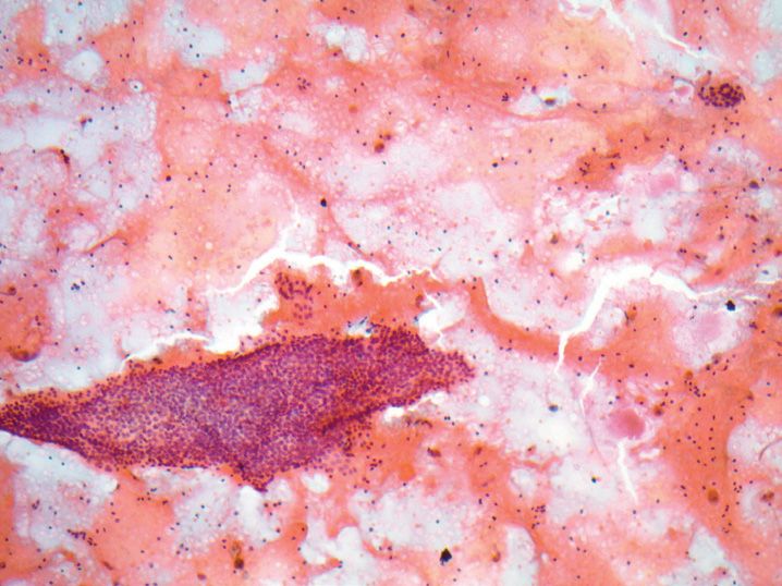
93. At endoscopy, esophageal brushings are performed, and show the following changes (see Figure 9-38). What clinical findings are most likely found in association with these changes?
(A) Circumferential stenosis caused by a friable mass
(B) Multiple small shallow ulcers in the distal esophagus
(C) Salmon-colored patches near the gastroesophageal junction
(D) Submucosal mass in the upper third of the esophagus
(E) White pseudomembranes that scrape off the epithelium
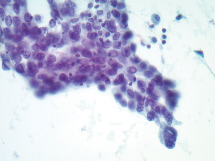
94. A 48-year-old woman undergoes thyroid fine-needle aspiration biopsy; she has multiple nodules, and the largest is aspirated (see Figure 9-39). The most appropriate interpretation for this aspirate, presuming that the figure provided is representative of the entire aspirate sample, is
(A) Atypia of undetermined significance
(B) Benign
(C) Malignant
(D) Suspicious for follicular neoplasm
(E) Suspicious for malignancy
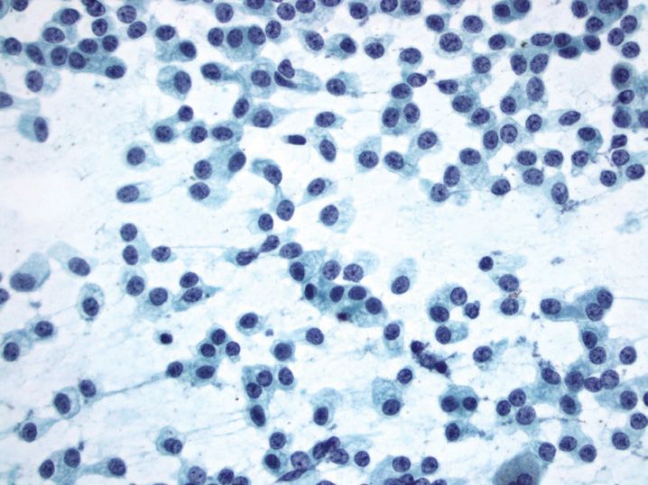
95. Transthoracic fine-needle aspiration biopsy of a peripherally located solitary, spiculated lung mass shows the following changes (see Figure 9-40). What is the most likely diagnosis?
(A) Carcinoid tumor
(B) Metastatic papillary thyroid carcinoma
(C) Reactive bronchial cells
(D) Squamous cell carcinoma
(E) Well-differentiated lung adenocarcinoma
96. A 45-year-old man with hematuria submits a urine sample for cytologic examination. Rare cells with large, dark nuclei and cytoplasmic golden pigment are seen (see Figure 9-41). What is the most likely origin of these cells?
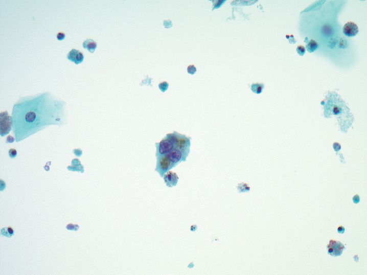
(A) Clear cell renal cell carcinoma
(B) High-grade urothelial carcinoma
(C) Seminal vesicle
(D) Squamous metaplasia of the trigone
(E) Urachal adenocarcinoma
97. A 75-year-old man who is a retired shipyard worker presents with dyspnea and a large left pleural effusion along with nodular thickening of the right pleural space. A portion of his right pleural effusion is submitted for cytologic examination, which shows large clusters of markedly atypical cells with scalloped borders and cytoplasmic windows. All of the following statements about this process are true except
(A) A distinctly different second population of benign mesothelial cells will be present in the effusion
(B) Electron microscopy will demonstrate long, thin microvilli (length-to-width ratio 15:1 or greater)
(C) Highly associated with asbestos exposure
(D) Immunohistochemical staining for calretinin will be positive in the neoplastic cells
(E) Low nuclear-to-cytoplasmic ratio is typical of this neoplasm
98. A 63-year-old woman undergoes fine-needle aspiration biopsy of a breast lesion associated with micro-calcifications on mammography. The aspirate smears demonstrate atypical epithelial cells with marked pleomorphism and hyperchromasia arranged as disorganized clusters and single atypical cells in a background of necrosis. At subsequent excision, which of the following lesions is this woman likely to have?
(A) Fibroadenoma
(B) High-grade ductal carcinoma in situ
(C) Infiltrating lobular carcinoma
(D) Intraductal papilloma
(E) Low-grade ductal carcinoma in situ
99. Fine-needle aspiration biopsy of an enlarged lymph node demonstrates necrosis, epithelioid histiocytes, and numerous neutrophils. Which of the following processes commonly results in this appearance?
(A) Cat scratch disease
(B) Chronic lymphocytic leukemia/small cell lymphoma
(C) Rosai–Dorfman disease
(D) Sarcoidosis
(E) Tuberculous lymphadenitis
100. All of the following are features of solid pseudopapillary tumor of the pancreas on cytology samples except
(A) Absence of nuclear staining for β-catenin
(B) Branching papillary-like structures with a vascular core
(C) Lack of cellular cohesion
(D) Myxoid or hyaline stroma
(E) Small monotonous cuboidal cells
101. Distinction between oncocytoma and an eosinophilic variant of chromophobe renal cell carcinoma on kidney fine-needle aspiration biopsy can be very difficult. Which of the following cytologic features favors a diagnosis of oncocytoma?
(A) Cytoplasmic clearing around the nucleus
(B) Diffuse cytoplasmic staining for Hale’s colloidal iron
(C) Marked variation in nuclear size
(D) Nuclear membrane irregularities
(E) Round-nested architecture on cell block preparation
102. A 5-year-old girl has a rapidly growing orbital mass. Fine-needle aspiration biopsy demonstrates a small round blue cell tumor with interspersed larger cells with elongated, dense cytoplasm, and eccentric nuclei. An immunohistochemical stain for desmin is positive in the neoplastic cells. What is the best diagnosis?
(A) Ewing sarcoma
(B) Fibrosarcoma
(C) Leiomyosarcoma
(D) Rhabdomyosarcoma
(E) Round cell liposarcoma
103. A parotid fine-needle aspiration biopsy demonstrates sheets of oncocyte-like cells in a background of naked nuclei. Which of the following stains, if positive, would favor a diagnosis of acinic cell carcinoma?
(A) Mucicarmine
(B) P63
(C) Periodic acid Schiff with diastase (PAS-D)
(D) Phosphotungstic acid hematoxylin (PTAH)
(E) S-100
104. A 52-year-old woman undergoes fine-needle aspiration biopsy of a thyroid nodule. The aspirate is interpreted as “suspicious for follicular neoplasm.” According to the 2007 Bethesda System for Reporting Thyroid Cytopathology, what is the risk of malignancy in this nodule?
(A) <3%
(B) 5–15%
(C) 15–30%
(D) 60–75%
(E) 97–99%
105. Prominent ciliocytophthoria, or detached tufts of cytoplasm containing a terminal bar and cilia, in a lung washing is associated with what viral infection?
(A) Adenovirus
(B) Cytomegalovirus
(C) Herpes simplex virus
(D) Measles virus
(E) Respiratory syncytial virus
106. A nipple discharge sample from a 32-year-old woman shows abundant foamy histiocytes and a background of proteinaceous debris. Which of the following conditions is this form of nipple discharge most likely to be associated with?
(A) Acute mastitis
(B) Galactorrhea
(C) Intraductal papilloma
(D) Invasive ductal carcinoma
(E) Tuberculous mastitis
107. Fine-needle aspiration biopsy of an enlarged lymph node demonstrates predominantly small mature lymphocytes, many of which are irregular or cleaved. Flow cytometry demonstrates a monotypic population of lymphocytes that are κ light chain restricted and express CD10. What is the best diagnosis for this aspirate?
(A) Burkitt lymphoma
(B) Diffuse large B-cell lymphoma
(C) Follicular lymphoma
Stay updated, free articles. Join our Telegram channel

Full access? Get Clinical Tree


