Cutaneous Plasmacytoma
Aaron Auerbach, MD, PhD
David Cassarino, MD, PhD
Key Facts
Terminology
Neoplasm of monoclonal plasma cells
Must 1st rule out plasmacytoid B-cell lymphoma
Clinical Issues
Skin is rare site for plasmacytomas; most occur in respiratory tract
Microscopic Pathology
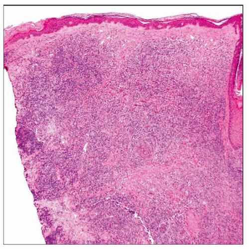
Diffuse or nodular collections of dermal plasma cells
Oval-shaped plasma cells with eccentric nucleus, “clock face” or “spoke wheel” chromatin without prominent nucleoli
Increased pink cytoplasm with perinuclear hof
Ancillary Tests
Usually CD38(+), CD138(+), pax-5(-), CD79a(+)
Flow cytometry underestimates number of plasma cells
Molecular shows clonal IgH rearrangement
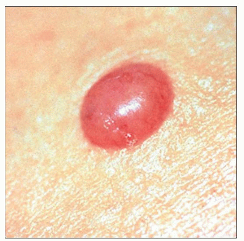
 A single red solitary plaque on the forearm is shown, which microscopically is a cutaneous plasmacytoma. |
TERMINOLOGY
Abbreviations
Cutaneous plasmacytoma (CP)
Synonyms
Extraosseous plasmacytoma of skin
Definitions
Neoplasm of monoclonal plasma cells in the skin
Must 1st exclude cutaneous involvement in multiple myeloma and plasmacytoid B-cell lymphoma such as primary cutaneous extranodal marginal zone lymphoma
CLINICAL ISSUES
Epidemiology
Incidence
Extremely rare
Only 3-5% of all plasma cell neoplasms
Many cases previously reported as primary cutaneous plasmacytomas would now be reclassified as extranodal marginal zone lymphomas
Rarely involve skin, more common in respiratory tract (80% of extraosseous plasmacytomas in oropharynx, nasopharynx, and nasal sinuses)
Rarely, may be seen as post-transplant lymphoproliferative disorder (PTLD)
Morphologically identical to other plasmacytomas, but PTLD plasmacytoma-like tumors are often EBV(+)
Age
Median age: 55 years
Gender
Mostly in men; male:female = 2:1
Stay updated, free articles. Join our Telegram channel

Full access? Get Clinical Tree


