Cutaneous Gamma-Delta T-cell Lymphoma
Aaron Auerbach, MD, PhD
Key Facts
Terminology
T-cell lymphoma of mature γδ cells
Separately classified from SPTCL in WHO (2008)
Clinical Issues
Hemophagocytic syndrome in 45%
Poor prognosis
Treated with multiagent chemotherapy
Macroscopic Features
Skin nodules with ulceration
Microscopic Pathology
3 patterns of disease: Epidermotropic, dermal, and subcutaneous
Malignant T cells rim around adipocytes
Prominent karyorrhexis/apoptosis and angioinvasion
Ancillary Tests
Immunohistochemistry: TCRδ1(+), βF1(−), CD56(+), CD4(−), CD8(−), EBER(−), cytotoxic markers (+)
T-cell receptor gene rearrangement
Top Differential Diagnoses
Subcutaneous panniculitis-like T-cell lymphoma
Panniculitis, but not in dermis or epidermis; lacks ulceration; TCRδ1(−), βF1(+)
Much better prognosis than CGDTCL
Peripheral T-cell lymphoma, not otherwise specified
Lupus profundus panniculitis
Similar inflammation in the subcutis in panniculitic pattern
Lobular panniculitis, but contains plasma cells and germinal centers, unlike CGDTCL
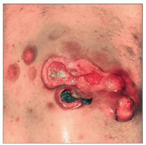 Cutaneous γδ T-cell lymphoma shows a large raised lesion with ulcer and satellite lesions. (Courtesy C. Sander, MD.) |
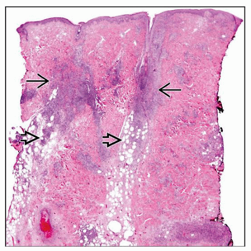 Primary cutaneous γδ T-cell lymphoma involves the epidermis, dermis  , and subcutaneous tissue , and subcutaneous tissue  with the most extensive disease in the dermis and subcutis. (Courtesy L. J. Medeiros, MD.) with the most extensive disease in the dermis and subcutis. (Courtesy L. J. Medeiros, MD.) |
TERMINOLOGY
Abbreviations
Cutaneous gamma-delta T-cell lymphoma (CGDTCL)
Synonyms
Subcutaneous panniculitis-like T-cell lymphoma with γδ cells
Definitions
T-cell lymphoma arising in the skin, which is composed of cytotoxic γδ T cells
Does not include subcutaneous panniculitis-like T-cell lymphoma composed of αβ cells
May encompass mucocutaneous γδ cell T-cell lymphoma, but further study is needed
ETIOLOGY/PATHOGENESIS
Immunosuppression or Dysregulation of T Cells
Found in many of the patients
Chronic Antigenic Stimulation
Speculative, but possibly involved in pathogenesis
Cell of Origin
γδ T cells
Involved with mucosal and epithelial immune system function
CLINICAL ISSUES
Epidemiology
Incidence
Rare tumor, < 100 cases reported in literature
< 1% of all cutaneous T-cell lymphomas
Age
Commonly adults
Gender
No gender preponderance
Site
Mostly extremities
Sometimes mucosal sites, where normal γδ T cells are found
Metastasis common
Spread to lungs, liver, kidneys, oral mucosa, and brain
Usually not in bone marrow, lymph node, or spleen
Presentation
1 or multiple skin lesions
Patches or plaques due to epidermal infiltrates
Tumors or nodules due to dermal infiltrates
± ulcerated epidermis
Hemophagocytic syndrome (HPS) may be present in 45% of cases
More often in subcutaneous lesions
Related to release of cytotoxic molecules
Laboratory Tests
Cytopenias
↑ liver function tests
Treatment
Adjuvant therapy
Multiagent chemotherapy ± radiotherapy
Poor response to allogenic stem cell transplant
Prognosis
Poor prognosis
5-year survival: ˜ 11%
Subcutaneous disease is a poor prognostic indicator
Better prognosis if only disease in dermis or epidermis
HPS is a poor prognostic indicator
MICROSCOPIC PATHOLOGY
Histologic Features
3 patterns of disease
Epidermotropic
Ranges from mild to marked
Can mimic mycosis fungoides or pagetoid reticulosis
Dermal
More dermal and epidermal involvement typically present than in subcutaneous panniculitis-like T-cell lymphoma
Subcutaneous
Lobules mostly involved
Septae less frequently involved and represents secondary spilling of T cells from lobules
Subcutis involvement can appear identical to subcutaneous panniculitis-like T-cell lymphoma of αβ cells, including rimming of fat cells
Often more than 1 pattern of disease in a patient
Different patterns of disease in a single biopsy or in different biopsies
Malignant T cells with nuclear atypia, hyperchromasia
Frequent necrosis/apoptosis and vascular invasion
Necrosis may be caused by released cytotoxic molecules
↑ reactive histiocytes
Vacuolated foamy cytoplasm from imbibed material/lipid
With erythrophagocytosis or cytophagocytosis
Cytologic Features
Medium to large T cells with coarse, hyperchromatic-staining chromatin, sometimes vesicular nuclei, and variably prominent nucleoli
ANCILLARY TESTS
Immunohistochemistry
T-cell antigens (CD2, CD3, CD5, CD7) positive
May lose 1 or more T-cell antigens
Most cases: CD4(−)/CD8(−)
Few cases: CD4(−)/CD8(+)
TCRδ1(+), βF1(−)
If TCRδ1 antibody is unavailable, βF1 can be used instead
Negative βF1 may serve to assume γδ cell origin
CD56 usually positive, unlike subcutaneous panniculitis-like T-cell lymphoma
Stay updated, free articles. Join our Telegram channel

Full access? Get Clinical Tree


