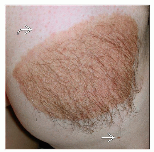
Congenital Melanocytic Nevi
Christine J. Ko, MD
Key Facts
Clinical Issues
Present at birth, by definition
However, some congenital melanocytic nevi may be “tardive” and not readily noticeable at birth
Categorized by size: < 1.5 cm = small; > 1.5 cm to < 20 cm = medium; > 20 cm = large
Risks, particularly for large congenital melanocytic nevus: ˜ 5% risk of developing cutaneous melanoma; at risk for neurocutaneous melanosis
Microscopic Pathology
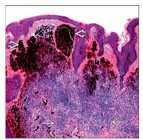
Melanocytes usually extend into lower reticular dermis and sometimes subcutaneous tissue with infiltration of arrector pili muscle, adnexal structures, nerves, and clustering around vessels
Cells become smaller with depth and less dense
Nodular proliferations (more common in large nevi): Based superficially in dermis, no epidermal involvement or necrosis
TERMINOLOGY
Synonyms
Giant/bathing suit/garment-type melanocytic nevus
CLINICAL ISSUES
Epidemiology
Incidence
1% of newborns
Age
Present at birth, by definition
However, some congenital melanocytic nevi may be “tardive”
Not readily noticeable at birth; becomes more obvious with age (generally by age 2)


