Condyloma Acuminatum (Genital Wart)
Elsa F. Velazquez, MD
Key Facts
Etiology/Pathogenesis
HPV-related lesion
Low-risk HPV serotypes (6, 11) predominate
Macroscopic Features
From flat to exophilic cauliflower-like lesions
Measuring from a few mm to several cm
Microscopic Pathology
Acanthosis with variable papillomatosis and prominent fibrovascular cores
Broad and sharply defined lower border
Conspicuous koilocytosis
Irregular wrinkled nuclei
Bi- and multinucleation
Perinuclear vacuolization
Top Differential Diagnoses
Verrucous carcinoma
Giant condyloma (Buschke-Löwenstein tumor)
Warty carcinoma
Papillary carcinoma, NOS
Papillomatosis of glans corona (pearly penile papules)
Bowenoid papulosis
Squamous cell carcinoma in situ (SCCis)
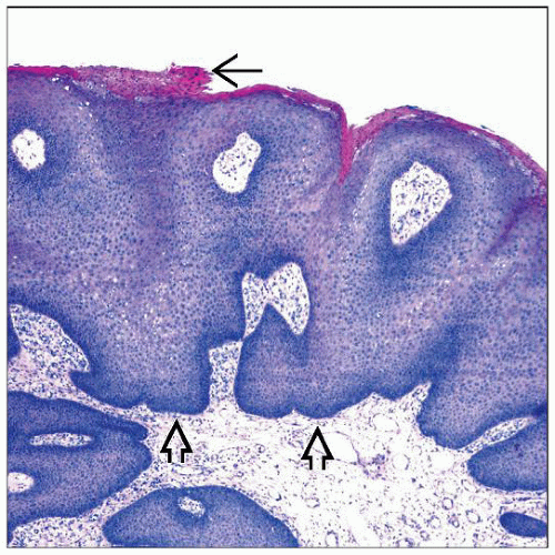 Condyloma acuminatum is characterized by marked acanthosis, slight (as in this example) to marked papillomatosis, and sharply demarcated bulbous base  . Note the focal parakeratosis . Note the focal parakeratosis  . . |
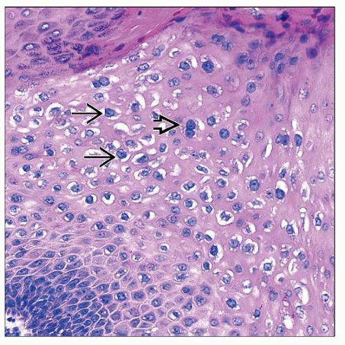 Koilocytes are characterized by clear perinuclear halos  and wrinkled and hyperchromatic nuclei. Binucleation is common and wrinkled and hyperchromatic nuclei. Binucleation is common  . The cytoplasm is eosinophilic. . The cytoplasm is eosinophilic. |
TERMINOLOGY
Synonyms
Genital wart
Definitions
Exophytic and verruciform nonmalignant epithelial lesions
ETIOLOGY/PATHOGENESIS
Infectious Agents
Caused by human papillomavirus (HPV)
Low-risk serotypes 6 and 11 (90% of cases)
Other serotypes include 16, 18, 30-32, 42-44, 51-55
> 1 serotype may be found in a lesion
CLINICAL ISSUES
Epidemiology
Incidence
Very common sexually transmitted disease
Age
Most frequent in young adults (2nd and 3rd decades of life)
Uncommon in children
Such cases should raise suspicion of sexual abuse
Site
Predilection for anogenital area
Males: Glans, prepuce, shaft
May extend to meatus
Females: Labia minora, interlabial sulcus, area around introitus
May extend into introitus
Both sexes: Perianal and more rarely oral cavity
Presentation
Soft fleshy verruciform plaques
Filiform lesions
Lesion in coronal sulcus and vulva may be bulkier and macerated
Some lesions may be almost flat and difficult to detect
Treatment
For small tumors: Cryosurgery, electrofulguration, laser ablation, and topical treatments
For medium-sized and large tumors: Surgical excision
Prognosis
Malignant transformation is rare in condyloma acuminatum
Patients with condyloma often have other sexually transmitted diseases
Stay updated, free articles. Join our Telegram channel

Full access? Get Clinical Tree


