Patient Story
A 21-year-old woman presents for her well-woman examination. She has been followed by her physician for many years and has no complaints. She has been sexually active for a little more than 2 years with one mutually monogamous partner. She does not smoke, has never had a sexually transmitted disease (STD), and uses oral contraceptive pills for contraception. On speculum examination, her cervix appears normal (Figure 88-1) and a Papanicolaou (Pap) test is performed.
Figure 88-1
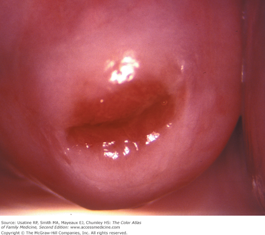
Normal cervix as seen through a colposcope without the application of vinegar. The red color around the os is produced by columnar cells and the lighter pink on the remainder of the cervix result from normal squamous cells. The presence of visible columnar cells outside the internal os is called ectropion and is a normal finding in young women and women on estrogen-containing contraception. The junction between the two cell types and colors is the squamocolumnar junction. (Courtesy of E.J. Mayeaux, Jr., MD.)
Introduction
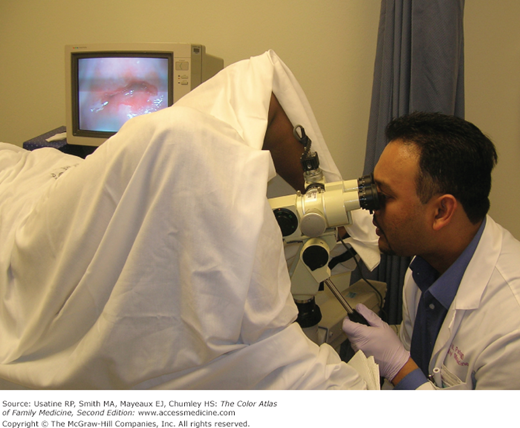
The colposcope is an optical instrument using light and magnification that helps distinguish dysplasia and cervical cancer from benign cervical findings (Figure 88-2). Colposcopy (colpo: vagina; scope: to look) literally means to look into the vagina. Primary indications for colposcopy include certain abnormal Pap results or an abnormal appearing cervix. Colposcopically directed biopsies have a higher yield than biopsies done without the benefit of a colposcope, thereby decreasing the risk of false-negative biopsies.
Synonyms
Epidemiology
- The Papanicolaou smear (Pap smear, Pap test) is a commonly employed screening test for dysplasia and cancer of the uterine cervix. More than 50 million Pap smears are performed each year in the United States.1 The Pap test is a cytologic examination of cells taken from the cervical transformation zone (Figures 88-3 and 88-4). Colposcopy is the diagnostic test to evaluate patients with an abnormal cervical cytologic smear or abnormal-appearing cervix.
- Nabothian cysts are common and benign and are considered a normal feature of the adult cervix. They may occur singly or multiple cysts may be found simultaneously (Figure 88-4).2
- Infections of the lower female genital tract are common and can produce a number of cervical epithelial changes (Figure 88-5).
- Cervical epithelial atrophy may occur in hypoestrogenic states and cause the cervix to appear pale and the squamocolumnar junction to retract into the cervical os (Figure 88-6).
- Endocervical polyps are the most common benign neoplasms of the uterine cervix (Figures 88-6 and 88-7) and are most commonly found incidentally during pelvic examination. They are most common in the fourth to sixth decades of life and usually are asymptomatic, but may cause vaginal discharge or postcoital spotting.3 Most polyps are benign with the incidence of malignancy being approximately 1 in 1000.4
Figure 88-3
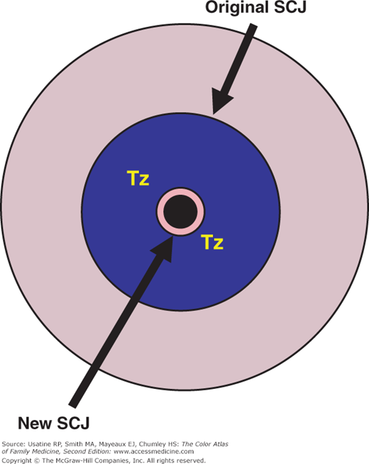
Schematic demonstration of the development of the transformation zone. The transformation zone extends from the original (prepubertal) squamocolumnar junction to the new (current) squamocolumnar junction. This transformation zone in the area at highest risk for cervical cancer. (Courtesy of E.J. Mayeaux, Jr., MD.)
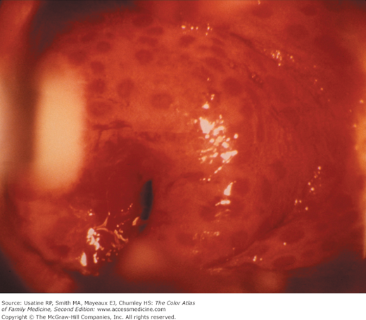
Figure 88-4
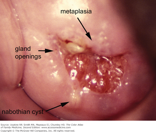
Normal transformation zone viewed with colposcopy. Note the squamocolumnar junction separating the red columnar epithelium from the pink squamous epithelium. Gland openings, nabothian cysts, and metaplasia are all part of this normal transformation zone. There is a white area of normal metaplasia on the anterior lip and a yellowish nabothian cyst on the posterior lip. (Courtesy of E.J. Mayeaux, Jr., MD.)