Chronic heart failure

21 Chronic heart failure


Key points


Chronic heart failure results from deficiency in the heart’s function as a pump, where the delivery of blood, and therefore oxygen and nutrients, becomes inadequate for the needs of the tissues. Chronic heart failure is a complex condition associated with a number of symptoms arising from defects in left ventricular filling and/or emptying, of which shortness of breath (exertional dyspnoea, orthopnoea and paroxysmal nocturnal dyspnoea), fatigue and ankle swelling are the most common. The symptoms of heart failure are due to inadequate tissue perfusion, venous congestion and disturbed water and electrolyte balance. Impairment of renal function, and the associated water retention, adds to the burden placed on the heart. In chronic heart failure, the physiological mechanisms that aim to maintain adequate tissue perfusion become counterproductive and contribute to the progressive nature of the condition.


Treatment is aimed at improving left ventricular function, controlling the secondary effects that lead to the occurrence of symptoms, and delaying disease progression. Drug therapy is indicated in all patients with heart failure to control symptoms (where present), improve quality of life and prolong survival. Patients with heart failure usually have their functional status assessed and categorised using the New York Heart Association (NYHA) classification system shown in Table 21.1.


Table 21.1 New York Heart Association (NYHA) classification of functional status of the patient with heart failure


I No symptoms with ordinary physical activity (such as walking or climbing stairs)
II Slight limitation with dyspnoea on moderate to severe exertion (climbing stairs or walking uphill)
III Marked limitation of activity, less than ordinary activity causes dyspnoea (restricting walking distance and limiting climbing to one flight of stairs)
IV Severe disability, dyspnoea at rest (unable to carry on physical activity without discomfort)


Aetiology


Heart failure may be a consequence of myocardial infarction, but as a chronic condition it is often gradual in onset with symptoms arising insidiously and without any specific cause over a number of years. The common underlying aetiologies in patients with heart failure are coronary artery disease and hypertension. The appropriate management of these predisposing conditions is also an important consideration in controlling heart failure in the community. Identifiable causes of heart failure include aortic stenosis, cardiomyopathy, mechanical defects such as cardiac valvular dysfunction, hyperthyroidism and severe anaemia. Conditions that place increased demands on the heart can create a shortfall in cardiac output and lead to intermittent exacerbation of symptoms. Symptoms of heart failure may occur as a consequence of hyperthyroidism, where the tissues place a greater metabolic demand, or severe anaemia, where there is an increased circulatory demand on the heart. Cardiac output may also be compromised by bradycardia or tachycardia, or by a sustained arrhythmia such as that experienced by patients in atrial fibrillation.


Atrial fibrillation often accompanies hyperthyroidism and mitral valve disease, where a rapid and irregular ventricular response can compromise cardiac efficiency. Improved management of the underlying causes, where appropriate, may alleviate the symptoms of heart failure, whereas the presence of mechanical defects may require the surgical insertion of prosthetic valve(s). While around 50% of patients with heart failure have significant left ventricular systolic dysfunction, the other half is comprised of patients who have either a normal or insignificantly reduced left ventricular ejection fraction (EF), although there is no consensus on the threshold for compromised EF and assessment of each patient relies mainly on clinical symptoms. These patients are referred to as having heart failure with preserved left ventricular ejection fraction (HFPEF). However, most of the available evidence from clinical trials regarding the pharmacological treatment of heart failure to date relates to those patients with heart failure due to left ventricular systolic dysfunction. Clinical symptomatic description of chronic heart failure is mild, moderate, or severe heart failure. ‘Mild’ is used for patients who are mobile with no important limitations of dyspnoea or fatigue, ‘severe’ for patients who are markedly symptomatic in terms of exercise intolerance and ‘moderate’ for those with restrictions in between. Trials tend to formalise these categories into NYHA Categories I–IV (Table 21.1).


Pathophysiology


In health, cardiac output at rest is approximately 5 L/min with a mean heart rate of 70 beats per minute and stroke volume of 70 mL. Since the filled ventricle has a normal volume of 130 mL, the fraction ejected is over 50% of the ventricular contents, with the remaining (residual) volume being approximately 60 mL. In left ventricular systolic dysfunction, the EF is reduced to below 45%, and symptoms are common when the fraction is below 35%, although some patients with a low EF can remain asymptomatic. When the EF falls below 10%, patients have the added risk of thrombus formation within the left ventricle and in most cases anticoagulation with warfarin is indicated.


Left ventricular systolic dysfunction can result from cardiac injury, such as myocardial infarction, or by exposure of the heart muscle to mechanical stress such as long-standing hypertension. This may result in defects in systolic contraction, diastolic relaxation, or both. Systolic dysfunction arises from impaired contractility, and is reflected in a low EF and cardiac dilation. Diastolic dysfunction arises from impairment of the filling process. Diastolic filling is affected by the rate of venous return, and normal filling requires active diastolic expansion of the ventricular volume. The tension on the ventricular wall at the end of diastole is called the preload, and is related to the volume of blood available to be pumped. That tension contributes to the degree of stretch on the myocardium. In diastolic dysfunction, there is impaired relaxation or reduced compliance of the left ventricle during diastole and, therefore, less additional blood is accommodated. In pure diastolic dysfunction, the EF can be normal but cardiac dilation is absent. Sustained diastolic dysfunction, which is a feature in a minority of patients with heart failure, may lead to systolic dysfunction associated with disease progression and left ventricular remodelling (structural changes and/or deterioration).


During systolic contraction, the tension on the ventricular wall is determined by the degree of resistance to outflow at the exit valve and that within the arterial tree, that is, the systemic vascular resistance. Arterial hypertension, aortic narrowing and disorders of the aortic valve increase the afterload on the heart by increasing the resistance against which the contraction of the ventricle must work. The result is an increased residual volume and consequently an increased preload as the ventricle overfills, and produces greater tension on the ventricular wall. In the normal heart, a compensatory increase in performance occurs as the stretched myocardium responds through an increased elastic recoil. In the failing heart, this property of cardiac muscle recoiling under stretch is diminished, with the consequence that the heart dilates abnormally to accommodate the increased ventricular load. With continued dilation of the heart the elastic recoil property can become much reduced. Failure of the heart to handle the increasing ventricular load leads to pulmonary and systemic venous congestion. At the same time, the increased tension on the ventricular wall in heart failure raises myocardial oxygen requirements, which increases the risk of an episode of myocardial ischaemia or arrhythmias.


The failing heart may show cardiac enlargement due to dilation, which is reversible with successful treatment. An irreversible increase in cardiac muscle mass, cardiac hypertrophy, occurs with progression of heart failure and is a consequence of long-standing hypertension. While hypertrophy may initially alleviate heart failure, the increased mass is pathologically significant because it ultimately increases the demands on the heart and oxygen consumption.


A reflex sympathetic discharge caused by the diminished tissue perfusion in heart failure exposes the heart to catecholamines where positive inotropic and chronotropic effects help to sustain cardiac output and produce a tachycardia. Arterial constriction diverts blood to the organs from the skin and gastro-intestinal tract but overall raises systemic vascular resistance and increases the afterload on the heart.


Reduced renal perfusion due to heart failure leads to increased renin release from the glomerulus in the kidney. Circulating renin raises blood pressure through the formation of angiotensin I and angiotensin II, a potent vasoconstrictor, and renin also prompts adrenal aldosterone release. Aldosterone retains salt and water at the distal renal tubule and so expands blood volume and increases preload. Arginine vasopressin released from the posterior pituitary in response to hypoperfusion adds to the systemic vasoconstriction and has an antidiuretic effect by retaining water at the renal collecting duct.


These secondary effects become increasingly detrimental to cardiac function as heart failure progresses, since the vasoconstriction adds to the afterload and the expanded blood volume adds to the preload. The expanded blood volume promotes the atrial myocytes to release a natural vasodilator, atrial natriuretic peptide (ANP), to attenuate the increased preload.


The compensatory mechanisms for the maintenance of the circulation eventually become overwhelmed and are ultimately highly counterproductive, leading to the emergence and progression of clinical signs and symptoms of heart failure. The long-term consequences are that the myocardium of the failing heart undergoes biochemical and histological changes that lead to remodelling of the left ventricle which further complicates disease progression. In those patients where the condition is severe and has progressed to an end stage, heart transplantation may be the only remaining treatment option.


Clinical manifestations


The reduced cardiac output, impaired oxygenation and diminished blood supply to muscles cause fatigue. Shortness of breath occurs on exertion (dyspnoea) or on lying (orthopnoea). When the patient lies down, the postural change causes abdominal pressure on the diaphragm which redistributes oedema to the lungs, leading to breathlessness. At night the pulmonary symptoms give rise to cough and an increase in urine production prompts micturition (nocturia), which adds to the sleep disturbance. The patient can be inclined to waken at night as gradual accumulation of fluid in the lungs may eventually provoke regular attacks of gasping (paroxysmal nocturnal dyspnoea). Characteristically the patient describes the need to sit or stand up to seek fresh air, and often describes a need to be propped up by three or more pillows to remedy the sleep disturbances that are due to fluid accumulation.


Patients with heart failure may appear pale and their hands cold and sweaty. Reduced blood supply to the brain and kidney can cause confusion and contribute to renal failure, respectively. Hepatomegaly occurs from congestion of the gastro-intestinal tract, which is accompanied by abdominal distension, anorexia, nausea and abdominal pain. Oedema affects the lungs, ankles and abdomen. Signs of oedema in the lungs include crepitations heard at the lung bases. In acute heart failure, symptoms of pulmonary oedema are prominent and may be life-threatening. The sputum may be frothy and tinged red from the leakage of fluid and blood from the capillaries. Severe dyspnoea may be complicated by cyanosis and shock. Table 21.2 presents the clinical manifestations of heart failure.


Table 21.2 Clinical manifestations of heart failure


Venous (congestion) Cardiac (cardiomegaly) Arterial (peripheral hypoperfusion)
Dyspnoea Dilation Fatigue
Oedema Tachycardia Pallor
Hypoxia Regurgitation Renal impairment
Hepatomegaly Cardiomyopathy Confusion
Raised venous pressure Ischaemia, arrhythmia Circulatory failure


Investigations


Patients with chronic heart failure are diagnosed and monitored on the basis of signs and symptoms from physical examination, history and an exercise tolerance test. On physical examination of the patient, a lateral and downward displacement of the apex beat can be identified as evidence of cardiac enlargement. Additional third and/or fourth heart sounds are typical of heart failure and arise from valvular dysfunction. Venous congestion can be demonstrated in the jugular vein of the upright reclining patient by an elevated jugular venous pressure (JVP), which reflects the central venous pressure. The JVP is measured by noting the visible distension above the sternum and may be accentuated in heart failure by the application of abdominal compression in the reclining patient. Confirmation of heart failure, however, should not be based on symptom assessment alone.


Echocardiography is important when investigating patients with a suspected diagnosis of heart failure. An echocardiogram allows visualisation of the heart in real time and will identify whether heart failure is due to systolic dysfunction, diastolic dysfunction or heart valve defects. With the provision of direct access echocardiography services to doctors in primary care, an increasing number of patients can now be quickly referred to confirm or exclude heart failure due to left ventricular systolic dysfunction or other structural abnormalities. Some reports suggest that between 50% and 75% of patients referred to direct access clinics may have normal left ventricular function, which has important implications for the selection of appropriate drug treatment. Table 21.3 shows a number of investigations that are routinely performed in the assessment of heart failure symptoms. The use of serum natriuretic peptide measurement in the diagnosis of patients with heart failure is currently limited by the lack of defined cut-off values and, therefore, measurements are only considered in combination with ECG/chest X-ray data prior to echocardiography.


Table 21.3 Investigations performed to confirm a diagnosis of heart failure


Investigation Comment
Blood test The following assessments are usually performed:


12-lead electrocardiogram A normal ECG usually excludes the presence of left ventricular systolic dysfunction. An abnormal ECG will require further investigation
Chest radiograph A chest radiograph (X-ray) is performed to look for an enlarged cardiac shadow and consolidation in the lungs
Echocardiography An echocardiogram is used to confirm the diagnosis of heart failure and any underlying causes, for example, valvular heart disease


Treatment of heart failure


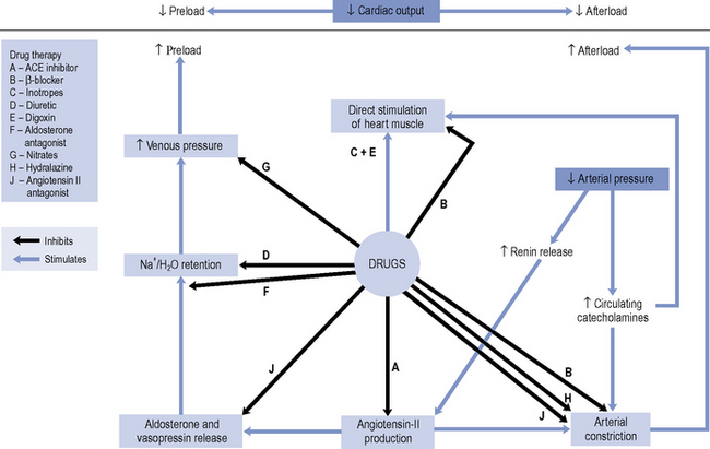
Until the 1980s, pharmacotherapy was driven by the aim to control symptoms, when diuretics and digoxin were the mainstay of treatment. While relieving the symptoms of heart failure remains decisive in improving a patient’s quality of life, a better understanding of the underlying pathophysiology has led to major advances in the pharmacological treatment of heart failure. With the introduction of angiotensin converting enzyme (ACE) inhibitors, β-blockers, angiotensin II receptor blockers (ARBs) and aldosterone antagonists, delaying disease progression and ultimately improving survival have become realistic goals of therapy. An outline of the site of action of the various drugs is schematically presented in Fig. 21.1.


In heart failure patients with co-morbid conditions known to contribute to heart failure, such as hyperthyroidism, anaemia, atrial fibrillation and valvular heart disease, attention must be given to ensuring these underlying contributing factors are well controlled. Patients with atrial fibrillation may be candidates for electrocardioversion. Tachycardia from atrial fibrillation usually requires control of the ventricular rate through suppression of atrioventricular node conduction. In patients with heart failure, the use of digoxin and/or β-blockers is recommended in such circumstances. In these patients, the use of either anticoagulant or antiplatelet agents is necessary and should be based on an assessment of stroke risk.


In patients with heart failure and preserved EF, diuretics are commonly used for symptom control and there is some limited evidence to suggest that ACE inhibitors can reduce hospitalisation. However, the use of all other agents of proven benefit in treating heart failure due to left ventricular systolic dysfunction are currently not supported by an evidence base.


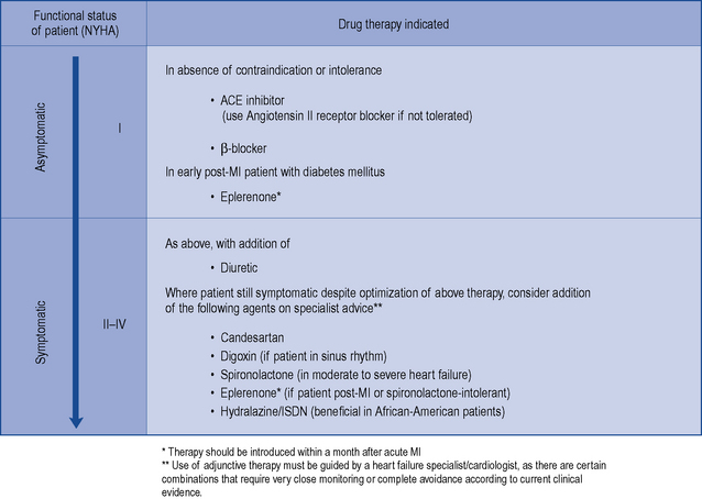
There is consensus that all patients with left ventricular systolic dysfunction should be treated with both an ACE inhibitor and a β-blocker in the absence of intolerance or contraindications. The evidence base for treatment clearly shows that use of an ACE inhibitor (or angiotensin receptor blocker) and β-blocker therapy in patients with heart failure due to left ventricular systolic dysfunction leads to an improvement in symptoms and reduction in mortality. There is some evidence to suggest that either agent can be initiated first, as both appear to be just as effective and well tolerated (CARMEN 2004, CIBIS III, 2005). Beneficial effects on morbidity and mortality have also been shown for the use of ARBs, aldosterone antagonists and hydralazine/nitrate combinations when used in the treatment of chronic heart failure. Digoxin has been shown to improve morbidity and reduce the number of hospital admissions in patients with heart failure, although its effect on mortality has not been demonstrated. Table 21.4 describes the treatment of acute heart failure in the hospital setting, while Fig. 21.2 highlights the possible treatment options for patients with chronic heart failure due to left ventricular systolic dysfunction.


Table 21.4 Treatment of acute heart failure due to left ventricular systolic dysfunction in patients requiring hospitalisation


Problem Drug therapy indicated
Anxiety Use of intravenous opiates to reduce anxiety and reduce preload through venodilation
Breathlessness High-flow oxygen (60–100%) may be required in conjunction with i.v. furosemide as either direct injection or 24-h infusion (5–10 mg/h).
Venodilation with i.v. GTN is also effective at doses titrated every 10–20 min against systolic BP ≤ 110 mmHg
Arrhythmia Digoxin useful in control of atrial fibrillation. Amiodarone is the drug of choice in ventricular arrhythmias
Expansion of blood volume following blood transfusion An elevation in preload, such as can occur acutely by expansion of blood volume after a transfusion, can exacerbate the degree of systolic dysfunction. Therefore, it is necessary to continue or increase diuretic dosage during this time
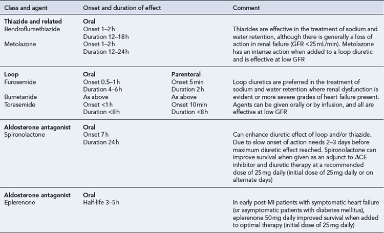

The selection of adjunctive therapy beyond the use of ACE inhibitor and β-blocker therapy is largely dependent on the nature of the patient and the preference of the heart failure specialist involved in the patient’s care. It is accepted that there is a limit as to how many agents any one patient can tolerate; therefore, the selection of drug therapy will probably be tailored to each individual patient, meaning that treatment plans will vary.


Diuretics


In chronic heart failure, diuretics are used to relieve pulmonary and peripheral oedema by increasing sodium and chloride excretion through blockade of sodium re-absorption in the renal tubule. Normally, in the proximal tubule, about 70% of sodium is reabsorbed along with water. In mild heart failure, either a thiazide or more often a loop diuretic is chosen depending on the severity of the symptoms experienced by the patient and the degree of diuresis required. Thiazides are described as ‘low-ceiling agents’ because maximum diuresis occurs at low doses, and they act mainly on the cortical diluting segment (the point of merger of the ascending limb with the distal renal tubule) at which 5–10% of sodium is normally removed. Although thiazides have some action at this site, they fail to produce a marked diuresis since a compensatory increase in sodium re-absorption occurs in the loop of Henle, and consequently thiazides are ineffective in patients with moderate-to-severe renal impairment (eGFR <30 mL/min) or persisting symptoms. Additionally, doses above the equivalent of bendroflumethiazide 5 mg have an increased risk of adverse metabolic effects with no additional symptomatic benefit. Thiazides are, therefore, now rarely used as sole diuretic therapy and are reserved for cases where the degree of fluid retention is very mild, renal function is not compromised or as an adjunct to loop diuretics (see below).


Loop diuretics are indicated in the majority of symptomatic patients and most patients will be prescribed one of either furosemide, bumetanide or torasemide in preference to a thiazide. These agents are known as ‘high-ceiling agents’ because their blockade of sodium re-absorption in the loop of Henle continues with increased dose. They have a shorter duration of action (average 4–6 h) compared to thiazides (average 12–24 h), and produce less hypokalaemia. In high doses, however, their intensity of action may produce hypovolaemia with risk of postural hypotension, worsening of symptoms and renal failure. In practice, high doses of furosemide (up to 500 mg/day) may be required to control oedema in patients with poor renal function. In the acute situation, doses of loop diuretics are titrated to produce a weight loss of 0.5–1 kg per day.


In longer term use, patients with heart failure frequently develop some resistance to the effects of loop diuretic due to a compensatory rebound in sodium retention. In this situation, a combination of thiazide and loop diuretics has been shown to have a synergistic effect, even in patients with reduced renal function. In the UK, metolazone is also used as an adjunct to augment the effects of loop diuretics. The potentially profound diuresis produced by such a combination poses serious risks, such as dehydration and hypotension, and patients who are prescribed metolazone in addition to an existing loop diuretic must be carefully monitored. In practice, patients with oedema treated with loop diuretics may best be treated using a degree of self-management. Some patients are instructed to make upward adjustment of loop diuretic dose or to add metolazone therapy on particular days, for example, when they self-record a gain of 2 kg or more in their body weight over a short period of time.


Diuretics also have a mild vasodilator effect that helps improve cardiac function and the intravenous use of loop diuretics reduces preload acutely by locally relieving pulmonary congestion before the onset of the diuretic effect. Effective diuretic therapy is demonstrated by normalisation of filling pressure. Therefore, continued elevation of the JVP suggests a need for more diuretic unless otherwise contraindicated. Intravenous furosemide must be administered at a rate not exceeding 4 mg/min to patients with renal failure, since it can cause ototoxicity when administered more rapidly.


Details of diuretic therapy used in left ventricular systolic dysfunction are summarised in Table 21.5.


ACE inhibitors


ACE inhibitors are indicated as first-line treatment for all grades of heart failure due to left ventricular systolic dysfunction, including those patients who are asymptomatic. These agents exert their effects by reducing both the preload and afterload on the heart, thereby increasing cardiac output.


ACE inhibitors act upon the renin–angiotensin–aldosterone system, and they reduce afterload by reducing the formation of angiotensin II, a potent vasoconstrictor in the arterial system. These drugs also have an indirect effect on sodium and water retention by inhibiting the release of aldosterone and vasopressin, thereby reducing venous congestion and preload. The increase in cardiac output leads to an improvement in renal perfusion, which further helps to alleviate oedema. ACE inhibitors also potentiate the vasodilator bradykinin and may intervene locally on ACE in cardiac and renal tissues.


ACE inhibitors are generally well tolerated by most patients and have been shown to improve the quality of life and survival in patients with mild-to-severe systolic dysfunction (CONSENSUS, 1987; CONSENSUS II, 1992; SOLVD-P, 1992; SOLVD-T, 1991; V-HeFT II, 1991), including those patients who have experienced a myocardial infarction (AIRE), 1993; SAVE, 1992; TRACE, 1995). When an ACE inhibitor is prescribed, it is important to ensure that the dose is started low and increased gradually, paying close attention to renal function and electrolyte balance. The dose should be titrated to achieve the target dose that has been associated with long-term benefits shown in clinical trials or (if not possible) the maximum tolerable dose. There is some evidence to suggest that high doses of ACE inhibitor are more effective than low doses in relation to reduction in mortality, although it is uncertain whether this is a general class effect (ATLAS, 1999). In clinical practice, it is possible that some patients may be treated with ACE inhibitors at doses below those used in clinical trials. As a consequence, actual outcomes in heart failure treatment may not be as good as expected from the trial findings.


The introduction of an ACE inhibitor may produce hypotension, which is most pronounced after the first dose and is sometimes severe. Patients at risk include those already on high doses of loop diuretics, where the diuretics cannot be stopped or reduced beforehand, and patients who may have a low-circulating fluid volume (due to dehydration) and an activated renin–angiotensin system. Hypotension can also occur where the ACE inhibitor has been initiated at too high a dose or where the dose has been increased too quickly after initiation. In the primary care setting, treatment must be started with a low dose which is usually administered at bedtime. In patients at particular risk of hypotension, a test dose of the shorter-acting agent captopril can be given to assess suitability for treatment before commencing long-term treatment with a preferred ACE inhibitor. Once it has been established that the ACE inhibitor can be initiated safely, the preferred option would be to switch to a longer acting agent with once- or twice-daily dosing, starting with a low dose which would be gradually titrated upwards to the recommended target (Table 21.6). Monitoring of fluid balance, blood biochemistry and blood pressure are essential safety checks during initiation and titration of ACE inhibitor therapy.


One of the most common adverse effects seen with ACE inhibitors is a dry cough and this is reported in at least 10% of patients. However, since a cough can occur naturally in patients with heart failure it is sometimes difficult to determine the true cause. ACE inhibitor therapy can also compromise renal function, although in patients in whom there is a reduction in renal perfusion due to worsening heart failure or hypovolaemia, renal dysfunction can also occur. Therefore, there are a number of instances where ACE inhibitor intolerance can be misdiagnosed in practice. Where ACE inhibitor intolerance is suspected, patients can usually be successfully rechallenged with an ACE inhibitor once their heart failure is more stable, although careful monitoring of the patient should be undertaken during initiation and subsequent dose titration. If the increase in the patient’s serum creatinine is >100% from baseline, the ACE inhibitor should be stopped, intolerance confirmed and specialist advice sought. Where the increase from baseline is 50–100%, the ACE inhibitor dose should be halved and serum creatinine concentration rechecked after 1–2 weeks. If renal function is stable and no cough or other adverse effects are reported, therapy should be continued. Where the problem persists, an alternative treatment option might be required, for example, ARB (similar benefits on morbidity and mortality, but there is a possibility of similar adverse effects on blood pressure and renal function) or hydralazine–nitrate combination.


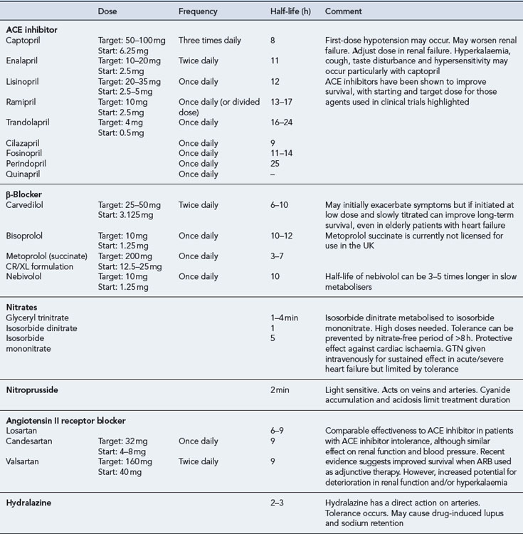
ACE inhibitors are potentially hazardous in patients with pre-existing renal disease, as blockade of the renin-angiotensin system may lead to reversible deterioration of renal function. In particular, ACE inhibitors are contraindicated in patients with bilateral renal artery stenosis, in whom the renin-angiotensin system is highly activated to maintain renal perfusion. Since most ACE inhibitors or their active metabolites rely on elimination via the kidney, the risk of other forms of dose-related toxicity is also increased in the presence of renal failure. Fosinopril, which is partially excreted by metabolism, may be the preferred agent in patients with renal failure. ACE inhibitors are also contraindicated in patients with severe aortic stenosis because their use can result in a markedly reduced cardiac output due to decreased filling pressure within the left ventricle. Table 21.6 summarises the activity and use of ACE inhibitors.


Angiotensin II receptor blockers


Although comparisons of ACE inhibitors and ARBs have shown similar benefits on morbidity and heart failure mortality, only ACE inhibitors have been shown to have positive effects on all cause mortality. ARBs should, therefore, not be used instead of ACE inhibitors, unless the patient experiences intolerable side effects.


The use of ARBs as an adjunct to ACE inhibitor and β-blocker therapy has been associated with significant reductions in cardiovascular events and hospitalisation rate (CHARM Added, 2003). Although this finding is encouraging, the impact on mortality alone remains inconsistent and there is no clear consensus on when to use an ARB as adjunctive therapy. In studies involving patients unable to tolerate an ACE inhibitor, ARBs have been shown to be comparable to ACE inhibitors in reducing the risk of cardiovascular death and rate of hospitalisation, and in the control of symptoms in heart failure patients (CHARM Alternative, 2003; Val-HeFT, 2002). Therefore, ARBs are recommended for use as an alternative to ACE inhibitor therapy where intolerance has been confirmed. It is important to note that in patients who have renal failure secondary to ACE inhibitors, switching to an ARB is of no theoretical or practical benefit, as similar adverse effects are likely.


A recent meta-analysis has raised concerns about a possible increase of cancer in people taking ARBs (Sipahi et al., 2010). Although the implications of this are unclear it adds weight to the recommendation that ACE inhibitors, not ARBs, should be the first-line agent when selecting a drug to act on the renin-angiotensin system.


β-Blockers


Formerly, β-blockers have been contraindicated in patients with heart failure. However, the sympathetic neurohormonal overactivity that occurs in response to the failing heart has been identified as a decisive factor in the progression of ventricular dysfunction. Consequently, β-blockers have been tested in a number of clinical trials. There is now substantial evidence that β-blockers reduce mortality among patients with mild-to-moderate symptomatic heart failure (ANZ Carvedilol, 1997; CAPRICORN, 2001; CIBIS II, 1999; MERIT-HF, 1999; US Carvedilol, 1996) and those with severe heart failure (COPERNICUS, 2001). This beneficial effect also extends to the elderly heart failure population (SENIORS, 2005).


The use of β-blockers is, therefore, recommended for all patients with heart failure due to left ventricular systolic dysfunction, irrespective of age and the degree of dysfunction. However, due to their negative inotropic effects, β-blockers should only be initiated when the patient’s condition is stable. There is insufficient evidence for a class effect to be assumed illustrated by the fact that in one trial, metoprolol tartrate was found to be inferior to carvedilol (COMET, 2003). Currently, nebivolol, bisoprolol and carvedilol are the only licensed β-blockers for the treatment of heart failure in the UK.


It is likely that patients will experience a worsening of symptoms during initiation of therapy and, therefore, patients are started on very low doses of β-blocker (e.g. carvedilol 3.125 mg daily) with careful titration occurring over a number of weeks or months with careful monitoring. The goal is to titrate the dose towards those used in clinical trials that have been associated with morbidity and mortality benefits (carvedilol 25–50 mg daily). Table 21.6 summarises the activity and use of β-blockers in heart failure.


Despite the demonstrated benefits, there is ongoing concern that certain subgroups of patients with heart failure continue to be undertreated with β-blockers. These groups include patients with chronic obstructive pulmonary disease (COPD), peripheral vascular disease, diabetes mellitus, erectile dysfunction and older adults. With the exception of patients with reversible pulmonary disease, who have typically been excluded from β-blocker trials (CIBIS II, 1999; MERIT-HF, 1999), there is now sufficient evidence to justify the use of β-blockers licensed for heart failure in these patients. In addition, a systematic review of trials on cardio-selective β-blockers found no clinically significant adverse respiratory effects in patients with reversible COPD, although it would be prudent to use these agents in such patients with caution and with appropriate monitoring in place (Salpeter S. et al., 2005).


Jun 18, 2016 | Posted by in PHARMACY | Comments Off on Chronic heart failure

Full access? Get Clinical Tree

Get Clinical Tree app for offline access