Choriocarcinoma
Key Facts
Terminology
Chorioepithelioma
Clinical Issues
Incidence
May represent the least common of all germ cell tumors in mediastinum
Represents < 5% of all germ cell tumors of mediastinum
Choriocarcinomas appear to be more common in 3rd and 4th decade of life
Choriocarcinomas are more common in men
Symptoms
Cough
Dyspnea
Gynecomastia
Chest pain
Superior vena cava syndrome
Laboratory findings
Increase HCG in serum
Image Findings
Anterior mediastinal mass
Macroscopic Features
Large bulky tumors
Extensive areas of hemorrhage and necrosis
Microscopic Pathology
Hemorrhage
Necrosis
Cytotrophoblast
Syncytiotrophoblast
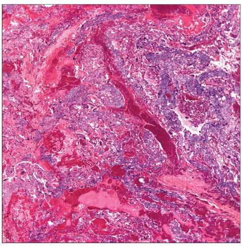 Primary mediastinal choriocarcinoma is seen with areas of hemorrhage admixed with neoplastic cellular proliferation without a specific growth pattern. |
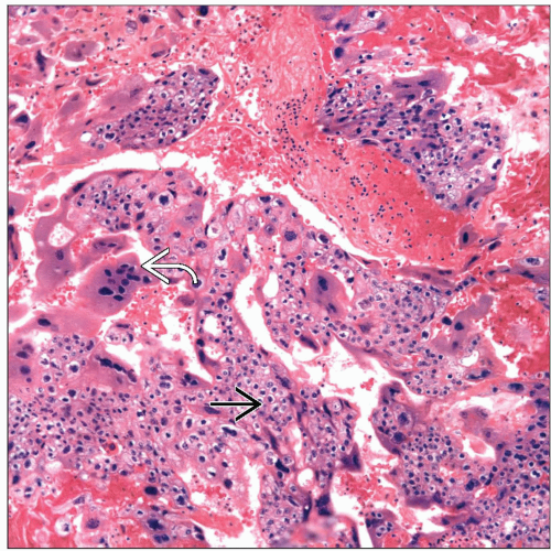 Choriocarcinoma with areas of hemorrhage admixed with a biphasic cellular proliferation is composed of large multinucleated cells (syncytiotrophoblast)  and round cells (cytotrophoblast) and round cells (cytotrophoblast)  . . |
TERMINOLOGY
Synonyms
Chorioepithelioma
Definitions
Malignant germ cell tumor
ETIOLOGY/PATHOGENESIS
Etiology
Misplaced germ cells in mediastinum
Unknown etiology
CLINICAL ISSUES
Epidemiology
Incidence
May represent the least common of all germ cell tumors in mediastinum
Accounts for < 5% of all germ cell tumors of mediastinum
Age
More common in 3rd and 4th decades of life
Gender
More common in males
Presentation
Cough
Dyspnea
Gynecomastia
Chest pain
Superior vena cava syndrome
Laboratory Tests
Increased hCG in serum
Treatment
Chemotherapy
Surgical debulking if necessary
Prognosis
Poor
Usually patients die within 12-18 months
Most patients at time of diagnosis already have widespread metastatic disease
IMAGE FINDINGS
General Features
Location
Anterior mediastinal mass
No pathognomonic features to separate choriocarcinoma from other nonseminomatous germ cell tumors
MACROSCOPIC FEATURES
General Features
Large bulky tumors
Extensive areas of hemorrhage and necrosis
Sections to Be Submitted
Due to presence of extensive necrosis and hemorrhage, extensive sampling is required
Size
Varies from a few cm to large tumors of > 10 cm in diameter
MICROSCOPIC PATHOLOGY
Histologic Features
Predominant Pattern/Injury Type
Hemorrhagic
Predominant Cell/Compartment Type
Germ, nonseminomatous
DIFFERENTIAL DIAGNOSIS
Pleomorphic Carcinoma (PC) of Lung Origin
Primary lung carcinomas may express hCG just like choriocarcinomas
Choriocarcinomas are usually tumors of younger patients in contrast to lung carcinoma
Choriocarcinomas are bulky anterior mediastinal masses, which may spread to lung
Lung carcinoma presents predominantly with a lung mass
Pleomorphic carcinoma is more common in older individuals
Pleomorphic carcinoma may show ectopic production of hCG
PC shows sarcomatous component with multinucleated giant cells
Choriocarcinoma shows presence of cyto- and syncytiotrophoblastic cells
Metastatic Choriocarcinoma of Gonadal Origin
Histology in both primary and metastatic disease is similar
Immunophenotype of both tumors is the same
In some cases of primary testicular choriocarcinoma, tumor may have undergone regression “burned-out”
It would be unusual for metastases to present with bulky anterior mediastinal mass
Clinical history would be very important
Thymic Carcinoma
Most thymic carcinomas do not show marked pleomorphism present in choriocarcinomas
Presence of cyto- and syncytiotrophoblastic components are not present in thymic carcinoma
Positive staining for hCG would be unusual in thymic carcinoma
Giant Cell Carcinoma
Usually shows malignant multinucleated giant cells not of syncytiotrophoblastic type of choriocarcinoma
Commonly shows cannibalism by giant cells
May show ectopic production of hCG
Does not show presence cytotrophoblastic component
Stay updated, free articles. Join our Telegram channel

Full access? Get Clinical Tree


