Chondroid Lipoma
Khin Thway, BSc, MBBS, FRCPath
Key Facts
Terminology
Uncommon benign neoplasm with features of embryonal fat and embryonal cartilage
Balanced translocation t(11;16)(q13; p12-13) in 2 cases, thought to be specific for lesion
Clinical Issues
Peak incidence in 3rd and 4th decades
Female preponderance
Occurs in superficial and deep locations
Most frequently affects proximal limbs and limb girdles
Microscopic Pathology
Nests and cords of rounded cells with granular eosinophilic cytoplasm or multivacuolated cytoplasm containing lipid
Prominent myxohyaline stroma
Variable amount of mature adipose tissue
S100 protein and CD68 positive
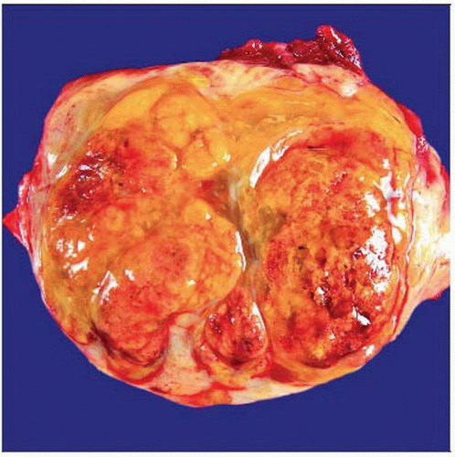 Gross photograph of chondroid lipoma shows a circumscribed lesion with a multilobulated, yellow to tan cut surface and prominent hemorrhage. |
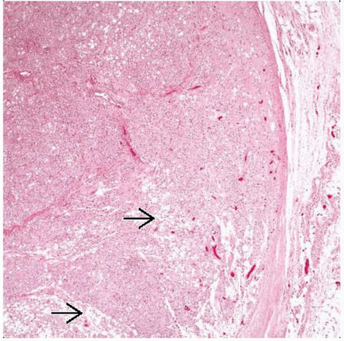 Hematoxylin & eosin shows a well-demarcated, encapsulated lesion within the subcutis. Sheets of cells with eosinophilic granular cytoplasm are admixed with mature adipocytes  . . |
TERMINOLOGY
Definitions
Uncommon benign neoplasm with features of embryonal fat and embryonal cartilage
Most frequently affects proximal limbs and limb girdles of adult women
Often mistaken for sarcomas
ETIOLOGY/PATHOGENESIS
Developmental Anomaly
Etiology unknown
CLINICAL ISSUES
Epidemiology
Age
Adults; peak incidence = 3rd and 4th decades
Gender
M:F = 1:4
Site
Predominantly proximal limbs and limb girdles
Trunk, head and neck (especially oral cavity)
Both superficial and deep locations
Presentation


