Chest radiograph showing right central venous line (solid white arrow) and ET tube (hollow white arrow) in correct positions. A left central line, which only reaches the brachiocephalic vein, requires repositioning (hollow black arrow). Note the left mid and lower zone consolidation (star).
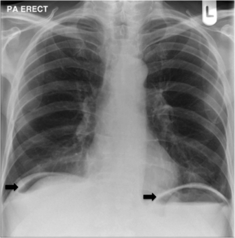
Chest radiograph showing free air below both hemi-diaphragms (arrows) from perforated abdominal viscus.
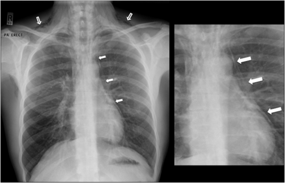
Chest radiograph shows surgical emphysema (hollow arrows) and air outlining the heart border and mediastinum (solid arrows), consistent with pneumomediastinum. This was caused by excessive vomiting (Boerhaave’s syndrome).
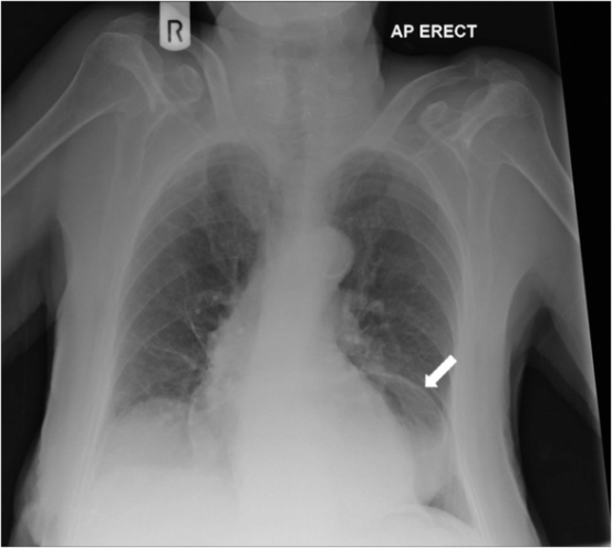
Chest radiograph demonstrating hiatus hernia: stomach present in the thorax.
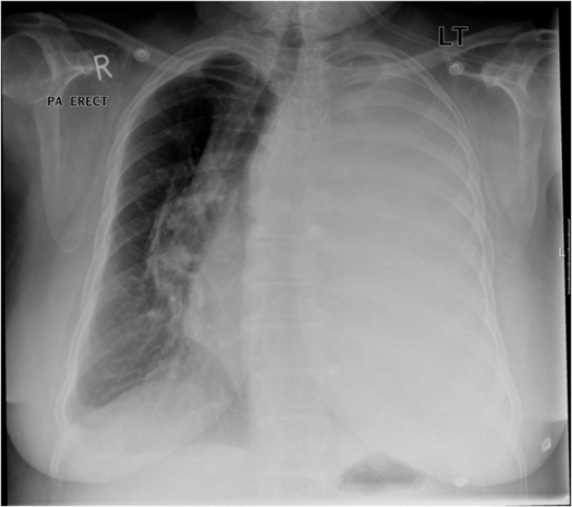
Chest radiograph demonstrating a very large left-sided pleural effusion causing midline shift of the mediastinum to the right. Note also tracheal deviation to the right
Stay updated, free articles. Join our Telegram channel

Full access? Get Clinical Tree


