Osteologic Overview
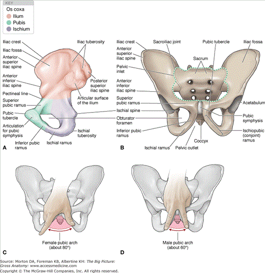
In the adult, the pelvis (os coxae) is formed by the fusion of three bones: ilium, ischium, and pubis (Figure 6-1A and B). The union of these three bones occurs at the acetabulum. The paired os coxae articulate posteriorly with the sacrum and anteriorly with the pubic symphysis.
The following structures are formed within the fused os coxa (Figure 6-1A–C):
- Acetabulum. A cup-shaped socket into which the ball-shaped head of the femur fits firmly.
- Obturator foramen. Covered by a flat sheet of connective tissue called the obturator membrane. A small opening located at the top of the membrane provides a route through which the obturator nerve, artery, and vein course.
- Greater sciatic notch. Located between the posterior inferior iliac spine and the ischial spine. The sacrospinous ligament converts the notch into the greater sciatic foramen, where the piriformis muscle, sciatic nerve, and pudendal neurovascular structures course.
- Lesser sciatic notch. Located between the ischial spine and the ischial tuberosity. The sacrotuberous ligament converts the notch into the lesser sciatic foramen.
- Pubic symphysis. Fibrocartilage connecting the two pubic bones in the anterior midline of the pelvis.
- Pelvic inlet. The superior aperture of the pelvis. The pelvic inlet is oval shaped and bounded by the ala of the sacrum, arcuate line, pubic bone, and symphysis pubis. The pelvic inlet is traversed by structures in the abdominal and pelvic cavities.
- Pelvic outlet. The inferior aperture of the pelvis. The pelvic outlet is a diamond-shaped opening formed by the pubic symphysis and sacrotuberous ligaments. Terminal parts of the vagina and the urinary and gastrointestinal tracts traverse the pelvic outlet. The perineum is inferior to the pelvic outlet.
The pelvic bone is formed by the fusion of three bones: ilium, ischium, and pubis.
- Iliac crest. Thickened superior rim.
- Iliac fossa. Concave surface on the anteromedial surface.
- Anterior superior iliac spine. Anterior termination of the iliac crest. Serves as an attachment site for the sartorius and tensor fascia lata muscles.
- Anterior inferior iliac spine. Serves as an attachment site for the rectus femoris muscle.
- Posterior superior iliac spine. Posterior termination of the iliac crest.
- Posterior inferior iliac spine. Forms the posterior border of the ala of the sacrum.
- Ischial tuberosity. A large protuberance on the inferior aspect of the ischium for attachment of the hamstring muscles and for supporting the body when sitting.
- Ischial spine. A pointed projection that separates the greater and lesser sciatic notches.
- Ischial ramus. A bony projection that joins with the inferior pubic ramus to form the ischiopubic ramus (conjoint ramus).
Stay updated, free articles. Join our Telegram channel

Full access? Get Clinical Tree


