Joints of the Digits and Fascia of the Foot
The foot is connected to the leg by the ankle (talocrural) joint, which is an articulation between the tibia, fibula, and talus. The foot consists of 7 tarsal bones, 5 metatarsal bones, and 14 phalanges. Motion at the digits for abduction and adduction is defined by an imaginary line along the long axis of the second digit, unlike the hand in which the long axis runs along the third digit. Each digit, with the exception of the great toe, consists of three phalanges (proximal, middle, and distal); the great toe has two phalanges (proximal and distal). The articulations between the bones of the foot create multiple joints. The muscles that move these joints are divided into two groups, intrinsic and extrinsic foot muscles. The intrinsic muscles originate and attach in the foot, whereas the extrinsic muscles originate in the leg and insert in the foot, creating motion at multiple joints.
The several bony articulations within the foot assist in accommodating uneven surfaces during weight-bearing activities. These motions of the foot are accomplished via the following joints (Figure 38-1):
- Metatarsophalangeal joints. Consist of articulations between the metatarsals and the proximal phalanges. The metatarsophalangeal joints allow flexion and extension and abduction and adduction.
- Interphalangeal joints. Consist of articulations between the phalanges, resulting in five proximal and four distal interphalangeal joints, which allow for flexion and extension.
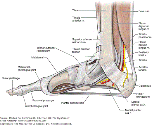
- Plantar aponeurosis. Radiates from the calcaneus bone toward the digits. The plantar aponeurosis is a very thick fascia that invests the muscles of the plantar surface of the foot.
- Superior extensor retinaculum. Attaches from the anterior border of the fibula to the tibia, proximal to the ankle joint. The superior extensor retinaculum holds the tendons of the tibialis anterior, extensor hallucis longus, extensor digitorum longus, and fibularis (peroneus) tertius muscles next to the structures of the anterior ankle during contraction.
- Inferior extensor retinaculum. A “Y-shaped” structure that attaches laterally to the superior surface of the calcaneus bone and courses medially to attach to the medial malleolus and the medial side of the plantar aponeurosis. The inferior extensor retinaculum serves to tether the tendons of the fibularis (peroneus) tertius, extensor digitorum longus, extensor hallucis longus, and anterior tibialis muscles.
- Flexor retinaculum. Attaches between the medial malleolus and calcaneus bones, forming the roof of the tarsal tunnel. The tendons of the tibialis posterior, flexor digitorum longus, and flexor hallucis longus muscles as well as tibial nerve and posterior tibial artery pass through the tarsal tunnel to the enter into the plantar surface of the foot.
- Fibular retinacula. Tethers the tendons of the fibularis (peroneus) longus and brevis muscles on the lateral side of the ankle as they course inferior to the lateral malleolus bone.
- Dorsal digital expansions. An aponeurosis covering the dorsum of the digits that attaches proximally to the middle phalanx (digits 2–5) or proximal phalanx (digit 1), via the central band, and distally to the distal phalanx, via the lateral bands. The extensor digitorum longus and brevis muscles and the extensor hallucis longus and brevis muscles attach proximally and centrally to the dorsal digital expansion. The lumbricals and the dorsal and plantar interossei attach on the free edges. Because of the attachment of the muscles and the location of the dorsal digital expansion, the small intrinsic muscles produce flexion at the metatarsophalangeal joint while extending the interphalangeal joints.
Muscles of the Foot
Muscles that act on the joints of the foot can either be extrinsic (originating outside the foot) or intrinsic (originating within the foot), and they may act on a single joint or multiple joints. The result is movement of multiple joints used to accommodate uneven surfaces or for activities such as running or jumping. The intrinsic muscles are discussed in this section, and the extrinsic muscles are discussed in Chapter 37.
The medial plantar or lateral plantar nerves originate from the tibial nerve and innervate the plantar muscles of the foot. The deep fibular nerve innervates the muscles on the dorsal side. These foot muscles are divided into four layers on the plantar surface and one group on the dorsal surface (Table 38-1).
Muscle | Proximal Attachment | Distal Attachment | Action | Innervation |
|---|---|---|---|---|
Layer 1 | ||||
Abductor digiti minimi | Calcaneal tuberosity | Lateral base of proximal phalanx 5 | Abduct and flex digit 5 | Lateral plantar n. (S1–S3) |
Flexor digitorum brevis | Both sides of middle phalanges digits 2–5 | Flex digits 2–5 | Medial plantar n. (S1–S2) | |
Abductor hallucis | Medial side of base of proximal phalanx digit 1 | Abduct and flex great toe | ||
Layer 2 | ||||


