Thigh
The bone between the hip and the knee is the femur. It is the longest and strongest bone in the body. The femur articulates proximally with the acetabulum and distally with the tibia and patella. The knee joint is formed by articulations of the femur, tibia, and patella. The knee joint enables flexion, extension, and minimal rotation of the femur and tibia. Also, it plays an important role in supporting the weight of the body during static positions and dynamic movement during gait.
The articulations between the femur, tibia, and patella form the knee joint and enable the following actions (Figure 36-1A):
- Flexion. Movement in the sagittal plane, decreasing the knee joint angle.
- Extension. Movement in the sagittal plane, increasing the knee joint angle.
- Medial rotation. Movement toward the midline in the transverse or axial plane.
- Lateral rotation. Movement away from the midline in the transverse or axial plane.
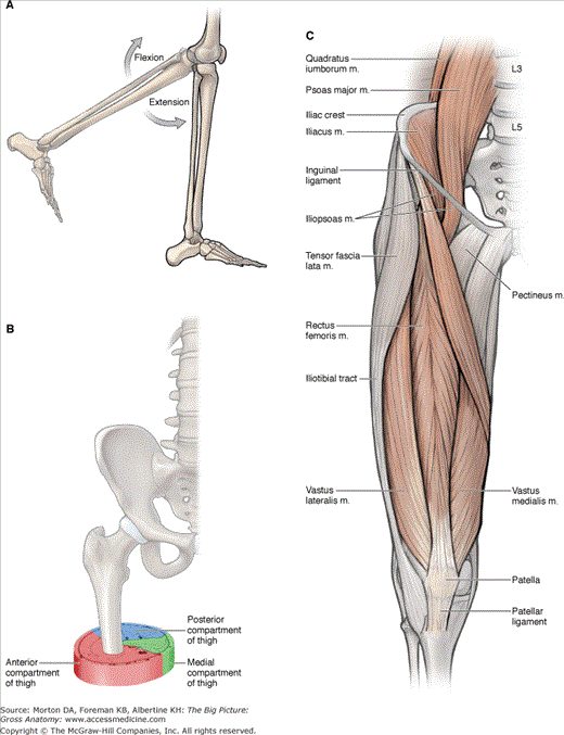
Muscles of the Thigh
The muscles of the thigh are divided by their fascial compartments (anterior, medial, and posterior) and may cross the hip or knee joint (Figure 36-1B). Identifying which joints the muscles cross and the side on which they cross can provide useful insight into the actions of these muscles (Table 36-1).
Muscle | Proximal Attachment | Distal Attachment | Action | Innervation |
|---|---|---|---|---|
Anterior compartment of the thigh | ||||
Psoas minor | T12–L1 vertebral bodies and discs | Pectin pubis | Lumbar spine flexion, posterior pelvic tilt | Anterior rami (L1) |
Psoas major | T12–L5 transverse processes, vertebral bodies and discs | Lesser trochanter of femur | Flexes and externally rotates thigh at hip joint; flexes trunk (psoas major) | Anterior rami (L1–L3) |
Iliacus | Iliac fossa | Femoral n. (L2, L3) | ||
Sartorius | Anterior superior iliac spine | Inferomedial to tibial tuberosity (pes anserinus) | Flexes thigh at hip joint and flexes leg at knee joint | Femoral n. (L2, L3) |
Rectus femoris | Anterior inferior iliac spine | Flexes thigh at hip joint and extends leg at knee joint | Femoral n. (L2–L4) | |
Vastus lateralis | Lateral part of intertrochanteric line, margin of greater trochanter, lateral margin of gluteal tuberosity, lateral lip of linea aspera | Quadriceps femoris tendon | Extends leg at knee joint | |
Vastus medialis | Medial part of intertrochanteric line, pectineal line, medial lip of linea aspera, medial supracondylar ridge | |||
Vastus intermedius | Femur: upper two-thirds of anterior and lateral surfaces | |||
Medial compartment of the thigh | ||||
Pectineus | Pectineal line | Oblique line extending from base of lesser trochanter to linea aspera on posterior surface of proximal femur | Adducts and flexes thigh at hip joint | Femoral n. (L2, L3) |
Adductor longus | Body of pubis | Linea aspera | Adducts and medially rotates thigh at hip joint | Obturator n. (anterior division) (L2–L4) |
Adductor brevis | Body of pubis and inferior pubic ramus | Obturator n. (anterior division) (L2, L3) | ||
Adductor magnus | Adductor part: ischiopubic ramus Hamstring part: ischial tuberosity | Adductor part: linea aspera Hamstring part: Adductor tubercle | Adducts and medially rotates thigh at hip joint | Adductor part: obturator n. (L2–L4) Hamstring part: tibial division of sciatic n. (L4) and obturator n. (L2, L3) |
Gracilis | Body and inferior ramus of pubic bone | Medial surface of proximal shaft of tibia (pes anserinus) | Adducts thigh at hip joint and flexes leg at knee joint | Obturator n. (L2, L3) |
Obturator externus | External surface of obturator membrane and adjacent bone | Trochanteric fossa | Laterally rotates hip | Obturator n. (posterior division) (L3, L4) |
Posterior compartment of the thigh | ||||
Semitendinosus | Ischial tuberosity | Medial surface of proximal tibia (pes anserinus) | Flexes leg at knee joint and extends thigh at hip joint; medially rotates thigh at hip joint and leg at knee joint | Tibial division of sciatic n. (L5–S2) |
Semimembranosus | Medial and posterior surface of medial tibial condyle | |||
Biceps femoris | Long head: ischial tuberosity Short head: lateral lip of linea aspera | Head of fibula | Knee flexion Hip extension Lateral rotation of hip and knee | Long head: tibial division of sciatic n. (L5–S2) Short head: common fibular division of sciatic n. (L5–S2) |
The muscles in the anterior compartment of the thigh are primarily flexors of the hip or extensors of the knee because of their anterior orientation (Figure 36-1C). The femoral nerve (L2–L4) innervates these muscles; however, each muscle does not necessarily receive each spinal nerve level between L2 and L4.
- Iliopsoas musculature. Originates from two muscles, the psoas major and iliacus muscles, which join to form a common tendon. The psoas major muscle attaches along vertebrae T12–L5, discs, and the iliacus within the iliac fossa. Both the psoas and iliacus muscles join together as they course deep to the inguinal ligament and insert onto the lesser trochanter of the femur. The main action of these muscles is to flex and laterally rotate the thigh at the hip joint. Innervation to the psoas major muscle is via the anterior rami of L1, L2, and L3, whereas innervation to the iliacus is through the femoral nerve (anterior rami of L2 and L3).
- Sartorius muscle. Attaches proximally to the anterior superior iliac spine. The distal insertion of the sartorius muscle is medial to the tibial tuberosity, contributing to the pes anserinus. Pes anserinus (“goose’s foot”) is a term used to describe the conjoined tendons of the sartorius, gracilis, and semitendinosus muscles; their common insertion is medial to the tibial tuberosity. The action of the sartorius muscle is to flex, abduct, and laterally rotate the thigh at the hip joint and flex the leg at the knee joint. The femoral nerve (L2 and L3) innervates this muscle.
- Quadriceps femoris muscle group. A four-headed muscle in the anterior compartment of the thigh and is a strong extensor muscle of the knee. There are four separate muscles in this group, each with distinct origins. However, all four parts of the quadriceps femoris muscle attach to the patella, via the quadriceps tendon, and then insert onto the tibial tuberosity. The femoral nerve (L2–L4) innervates the quadriceps femoris muscle group. The four separate muscles are as follows:
- Rectus femoris muscle. Attaches on the anterior inferior iliac spine and to the quadriceps femoris tendon. The rectus femoris muscle flexes the thigh at the hip joint and extends the leg at the knee joint.
- Vastus lateralis muscle. Attaches proximally at the intertrochanteric line and the lateral lip of the linea aspera; distally, the muscle attaches to the quadriceps femoris tendon. The vastus lateralis muscle extends the leg at the knee joint.
- Vastus medialis muscle. Attaches proximally at the intertrochanteric line and the lateral lip of the linea aspera; distally, the muscle attaches to the quadriceps femoris tendon. The vastus medialis muscle extends the leg at the knee joint.
- Vastus intermedius muscle. Attaches proximally along the anterior and lateral surfaces of the upper two-thirds of the femoral shaft; distally, the muscle attaches to the quadriceps femoris tendon. The vastus intermedius muscle extends the leg at the knee joint.
- Rectus femoris muscle. Attaches on the anterior inferior iliac spine and to the quadriceps femoris tendon. The rectus femoris muscle flexes the thigh at the hip joint and extends the leg at the knee joint.
The muscles in the medial compartment of the thigh are primarily adductors of the hip because of their medial orientation. The obturator nerve (L2–L4) innervates most of the muscles in the medial compartment of the thigh. However, each muscle does not necessarily receive each spinal nerve level between L2 and L4 (Figure 36-2A and B).
- Pectineus muscle. Attaches to the pectineal line of the pubis and the posterior surface of the proximal femur. The pectineus muscle adducts and flexes the thigh at the hip joint. The femoral nerve (L2 and L3) innervates this muscle, with occasional branches from the obturator nerve.
- Adductor longus muscle. Attaches proximally to the body of the pubis; distally, the muscle attaches on the linea aspera. The adductor longus muscle adducts and medially rotates the thigh at the hip joint. The obturator nerve (L2–L4) innervates this muscle.
- Adductor magnus muscle. Consists of an adductor part and a hamstring part

Stay updated, free articles. Join our Telegram channel

Full access? Get Clinical Tree


