Organization of the Fascia of the Hand
The fascia of the hand is continuous with the fascia of the forearm (antebrachium). In the hand, the fascia varies in thickness and divides the hand into five separate compartments that correspond with the five digits and have similar blood supply, innervation, and actions.
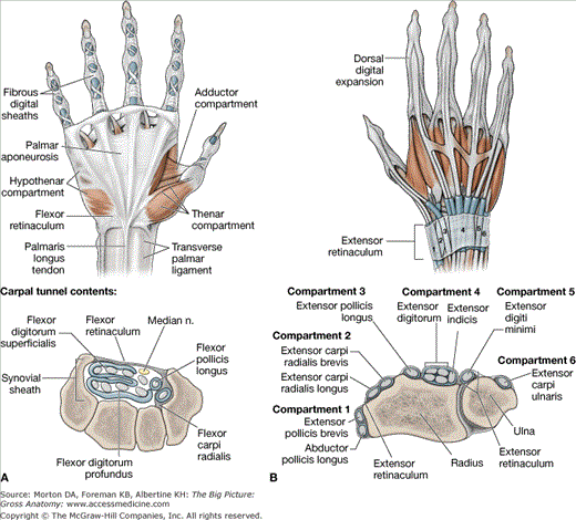
- Palmar aponeurosis. Located over the palm of the hand and covers the flexor tendons and deeper structures of the hand. The palmar aponeurosis extends distally and becomes continuous with the fibrous digital sheaths.
- Fibrous digital sheaths. Form a tunnel that encloses the flexor tendons of digits 2 to 5 and the tendon of the flexor pollicis longus muscle and their associated synovial sheaths.
- Flexor retinaculum (transverse carpal ligament). Forms a roof over the concavity created by the carpal bones, forming a tunnel (i.e., the carpal tunnel). The median nerve and the tendons of the flexor digitorum superficialis, flexor digitorum profundus, and flexor pollicis longus muscles, and their associated synovial sheaths, pass through this tunnel. The flexor retinaculum anchors medially to the pisiform and the hook of the hamate. Laterally, the flexor retinaculum is anchored to the scaphoid and trapezium.
- Transverse palmar ligament. Continuous with the extensor retinaculum from the dorsal side of the wrist and wraps around, anteriorly, to form a fascial band around the flexor tendons. This ligament should not be confused with the flexor retinaculum, which is located deeper to the transverse palmar ligament.
The fascial layers divide the palmar side of the hand into the following five compartments (Figure 33-1A):
- Thenar compartment. Contains three muscles that act on digit 1 (thumb).
- Hypothenar compartment. Contains three muscles that act on digit 5.
- Central compartment. Located between the thenar and hypothenar compartments and contains the flexor tendons and the lumbrical muscles.
- Adductor compartment. Contains the adductor pollicis muscle.
- Interosseous compartment. Located between the metacarpals and contains the dorsal and palmar interossei muscles.
- Extensor retinaculum. Continuous with the fascia of the forearm and attached laterally to the radius and medially to the triquetrum and pisiform bones. The extensor retinaculum works to retain the tendons that are near the bone while allowing proximal and distal gliding of the tendons (Figure 33-1B).
- Dorsal digital expansions. An aponeurosis covering the dorsum of the digits and attaches distal to the distal phalanx. Proximally and centrally, the extensor digitorum, extensor digiti minimi, extensor indicis, and extensor pollicis brevis muscles attach to the dorsal digital expansion. Laterally, the lumbricals and the dorsal and palmar interossei muscles attach. The small intrinsic muscles that attach laterally are responsible for delicate finger movements that would not be possible with the extensor digitorum, flexor digitorum superficialis, and profundus muscles alone. Because of the attachment of the muscles and the location of the hood, the small intrinsic muscles will produce flexion at the metacarpophalangeal joint while extending the interphalangeal joints.
The extensor retinaculum of the hand divides the dorsum of the wrist into the following six compartments:
- Compartment 1. Contains the abductor pollicis longus and extensor pollicis brevis muscles.
- Compartment 2. Contains the extensor carpi radialis longus and brevis muscles.
- Compartment 3. Contains the extensor pollicis longus muscles.
- Compartment 4. Contains the extensor digitorum and extensor indicis muscles.
- Compartment 5. Contains the extensor digiti minimi muscles.
- Compartment 6. Contains the extensor carpi ulnaris muscles.
Actions of the Fingers and Thumb
The hand consists of five digits (four fingers and a thumb). The thumb is considered digit 1; index finger is digit 2; middle finger is digit 3, ring finger is digit 4, and the little finger is digit 5. There are 19 bones and 19 joints in the hand distal to the carpal bones. Each digit has carpometacarpal, metacarpophalangeal, and interphalangeal joints.
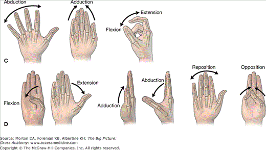
The finger joints and associated movements are as follows (Figure 33-1C):
- Carpometacarpal joints. Sliding joints that allow for gliding and rotation.
- Metacarpophalangeal joints. Condylar joints that allow for flexion and extension as well as abduction and adduction. Movements of abduction and adduction are described in relation to digit 3 (the middle finger). All movements away from digit 3 are considered abduction, and movements toward digit 3 are considered adduction. Rotation is limited because of the collateral ligaments.
- Interphalangeal joints. Hinge joints that allow for flexion and extension. There is a proximal interphalangeal joint and a distal interphalangeal joint for digits 2 to 5. They are often referred to as PIP and DIP, respectively.
The thumb joints and associated movements are as follows (Figure 33-1D):
- Carpometacarpal joint. Saddle joint that allows for opposition and reposition.
- Metacarpophalangeal joint. Hinge joint that allows for flexion and extension.
- Interphalangeal joint. Hinge joint that allows for flexion and extension. There is only one interphalangeal joint for digit 1 (the thumb).
The thumb is rotated 90 degrees to digits 2 to 5. Therefore, abduction and adduction occur in the sagittal plane, and flexion and extension occur in the coronal plane.
Muscles of the Hand
Muscles that act on the joints of the hand can be either extrinsic (originating outside the hand) or intrinsic (originating within the hand), and they may act on a single joint or on multiple joints. The result is movement of multiple joints for activities such as functional grasping or writing (Table 33-1).
Muscle | Proximal Attachment | Distal Attachment | Action | Innervation |
|---|---|---|---|---|
Palmaris brevis | Palmar aponeurosis and flexor retinaculum | Dermis on the ulnar side of hand | Tenses the skin over the hypothenar muscles | Ulnar n., superficial branch (C8–T1) |
Thenar muscles | ||||
Abductor pollicis brevis | Flexor retinaculum, scaphoid, and trapezium | Proximal phalanx of digit 1 | Abduction of thumb | Median n., recurrent branch (C8–T1) |
Flexor pollicis brevis | Deep head: trapezium and flexor retinaculum Superficial head: trapezoid and capitate | Flexion of digit 1 (metacarpophalangeal joint) | ||
Opponens pollicis | Trapezium and flexor retinaculum | Metacarpal 1 | Medial rotation of thumb and flexion of metacarpal of digit 1 | |
Adductor compartment | ||||
Adductor pollicis | Oblique head: metacarpals 2 and 3 and capitate Transverse head: metacarpal 3 | Proximal phalanx of digit 1 | Adduction of thumb | Ulnar n., deep branch (C8–T1) |
Hypothenar muscles | ||||
Abductor digit minimi | Pisiform bone, pisohamate ligament, and tendon of flexor carpi ulnaris | Proximal phalanx of digit 5 | Abduction of digit 5 | Ulnar n., deep branch (C8–T1) |
Flexor digiti minimi brevis | Hook of hamate and flexor retinaculum | Flexion of metacarpophalangeal joint of digit 5 | ||
Opponens digiti minimi | Metacarpal 5 | Lateral rotation of metacarpal 5 | ||
Central compartment | ||||
Lumbricals 1 and 2 | Lateral two tendons of flexor digitorum profundus | Lateral sides of dorsal digital expansions for digits 2–5 |
||


