Arm
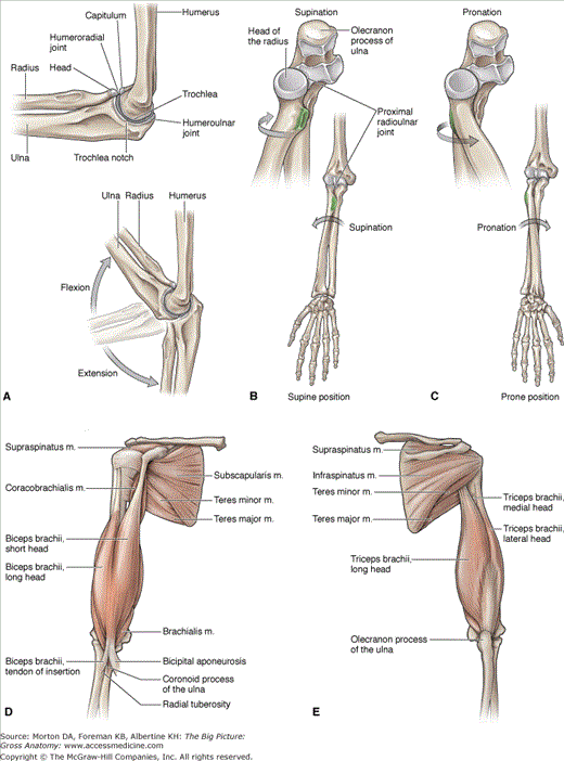
The arm (brachium) consists of the humerus, which articulates distally with the forearm (antebrachium) through the elbow complex. The elbow complex consists of three bones: humerus, ulna, and radius. The articulations of these bones result in three separate joints that share a common synovial cavity, enabling the forearm to flex, extend, pronate, and supinate on the humerus.
The articulations of the humerus, radius, and ulna in the elbow result in the following actions:
- Flexion and extension (Figure 31-1A)
- Humeroulnar joint. Articulation between the trochlear notch of the ulna and the trochlea of the humerus.
- Humeroradial joint. Articulation between the head of the radius and the capitulum of the humerus.
- Humeroulnar joint. Articulation between the trochlear notch of the ulna and the trochlea of the humerus.
- Pronation and supination (Figure 31-1B and C)
- Proximal radioulnar joint. Articulation between the head of the radius and the radial notch of the ulna.
Muscles of the Arm
The muscles of the arm are divided by their fascial compartments (anterior and posterior), and may cross one or more joints. Identifying the joints that the muscles cross and the side on which they cross can provide useful insight into the actions of these muscles (Table 31-1).
Muscle | Proximal Attachment | Distal Attachment | Action | Innervation |
|---|---|---|---|---|
Anterior compartment of the arm | ||||
Biceps brachii | Long head: supraglenoid tubercle Short head: coracoid process | Radial tuberosity | Flexion of shoulder and flexion and supination of elbow | Musculocutaneous n. (C5–C6) |
Brachialis | Distal anterior surface of humerus | Coronoid process and tuberosity of ulna | Flexion of the elbow | Musculocutaneous n. (C5–C6) & radial n. (C7) |
Coracobrachialis | Coracoid process of scapula | Medial, midshaft surface of humerus | Flexion of shoulder | Musculocutaneous n. (C5–C7) |
Posterior compartment of the arm | ||||
Triceps brachii | Long head: infraglenoid tubercle Lateral head: posterior humerus Medial head: posterior humerus | Olecranon process of ulna | Extension of shoulder and elbow | Radial n. (C6–C8) |
The muscles in the anterior compartment of the arm are primarily flexors (of the shoulder or elbow or both) because of their anterior orientation (Figure 31-1D). The musculocutaneous nerve (C5–C7) innervates the muscles in the anterior compartment of the arm. However, each muscle does not necessarily receive each spinal nerve level between C5 and C7. The following muscles are located in the anterior compartment of the arm:
- Coracobrachialis muscle. Attaches between the coracoid process of the scapula and the midshaft of the humerus. The coracobrachialis muscle crosses anteriorly to the glenohumeral joint and, therefore, contributes to shoulder flexion. It receives its innervation from the musculocutaneous nerve (C5–C7) and its blood supply via branches of the axillary artery.
- Brachialis muscle. Attaches between the anterior aspect of the humerus and the coronoid process and the tuberosity of the ulna, crossing the anterior elbow joint. The brachialis muscle acts on the ulna (humeroulnar joint), and therefore, it produces flexion of the elbow. As with the other muscles in the anterior compartment of the arm, the musculocutaneous nerve (C5–C6) provides innervation. However, the radial nerve (C7) innervates a small, lateral portion of the muscle. Blood is supplied to the muscle by branches from the brachial artery.
- Biceps brachii muscle. Consists of two heads that attach to the supraglenoid tubercle (long head) and the coracoid process (short head). The biceps brachii muscle converges to insert on the radial tuberosity. The biceps brachii crosses anterior to the glenohumeral joint and the elbow, primarily producing flexion

Stay updated, free articles. Join our Telegram channel

Full access? Get Clinical Tree


