Shoulder Complex
The combined joints connecting the scapula (scapulothoracic joint), clavicle (sternoclavicular and acromioclavicular joints), and humerus (glenohumeral joint) form the shoulder complex and anchor the upper limb to the trunk. The only boney stability of the upper limb to the trunk is through the connection between the clavicle and the sternum. The remaining stability of the shoulder complex depends on muscles, and as a result, the shoulder complex has a wide range of motion.
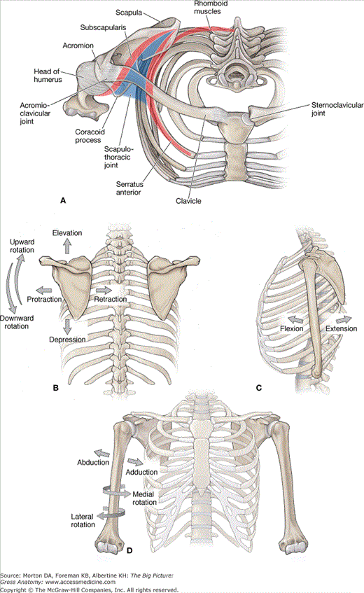
To best understand the actions of the scapula, it is important to understand the scapulothoracic, acromioclavicular, and sternoclavicular joints (Figure 30-1A).
- Scapulothoracic joint. Formed by the articulation of the scapula with the thoracic wall through the scapular muscles, including the trapezius and the serratus anterior muscles. The scapulothoracic joint is not considered a true anatomic joint; as such, it is frequently referred to as a “pseudo joint” because it does not contain the typical joint characteristics (e.g., synovial fluid and cartilage).
- Acromioclavicular joint. A synovial joint formed by the articulations of the scapula (acromion) and the clavicle.
- Sternoclavicular joint. A synovial joint formed by the articulations between the clavicle and sternum.
The scapulothoracic, sternoclavicular, and acromioclavicular joints are interdependent. For example, if motion occurs at one joint (e.g., the scapula elevates), the movement will directly affect the other two joints. Therefore, the motions produced frequently involve more than a single joint. Although the scapular movements include the scapulothoracic, acromioclavicular, and sternoclavicular joints, we will refer only to the scapula in the following text.
The following terms describe the movements of the scapula (Figure 30-1B):
- Protraction. Anterior movement of the scapula on the thoracic wall (e.g., reaching in front of the body).
- Retraction. Posterior movement of the scapula on the thoracic wall (e.g., squeezing the shoulder blades together).
- Elevation. Raising the entire scapula in a superior direction without rotation.
- Depression. Lowering the entire scapula in an inferior direction without rotation.
- Upward rotation. Named according to the upward rotation and direction that the glenoid fossa faces.
- Downward rotation. Named according to the downward rotation and direction that the glenoid fossa faces.
The glenohumeral joint is a synovial, ball-and-socket joint. The “ball” is the head of the humerus, and the “socket” is the glenoid fossa of the scapula. The glenohumeral joint is considered to be the most mobile joint in the body and produces the following actions (Figure 30-1C and D):
- Flexion. Movement anterior in the sagittal plane.
- Extension. Movement posterior in the sagittal plane.
- Abduction. Movement away from the body in the frontal plane.
- Adduction. Movement toward the body in the frontal plane.
- Medial rotation. Movement toward the body in the transverse or axial plane.
- Lateral rotation. Movement away from the body in the transverse or axial plane.
- Circumduction. A combination of glenohumeral joint motions that produce a circular motion.
Muscles of the Shoulder Complex
The musculature of the scapulothoracic joint is responsible primarily for the stability of the scapula to provide a stable base for the muscles acting on the glenohumeral joint. In other words, the upper limb must have proximal stability to have distal mobility. In addition, the scapulothoracic joint works in conjunction with the glenohumeral joint to produce movements of the shoulder. For example, the available range of motion for shoulder abduction is 180 degrees. This motion is produced by approximately 120 degrees from abduction at the glenohumeral joint and by approximately 60 degrees of upward rotation from the scapulothoracic joint.
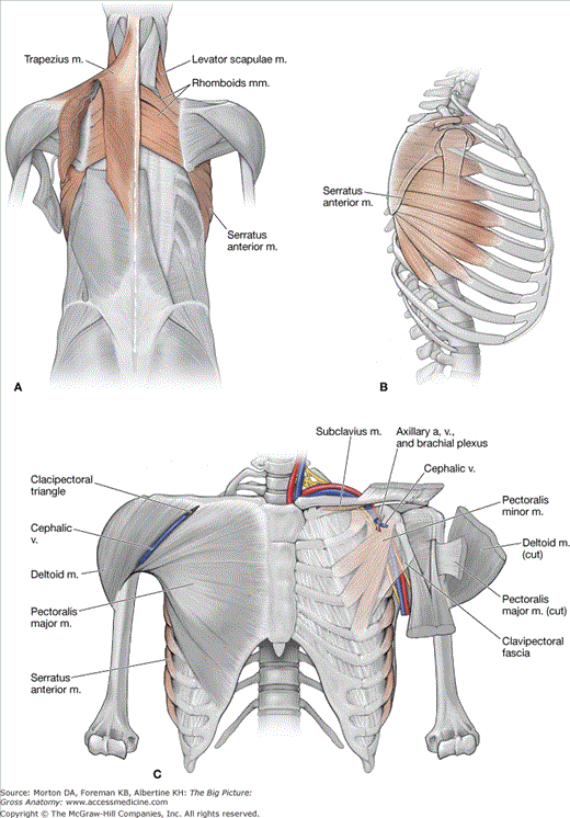
The following are muscles of the scapula (Figure 30-2A–C and Table 30–1):
- Trapezius muscle. Attaches to the occipital bone, nuchal ligament, spinous processes of C7–T12, spine of the scapula, acromion, and the clavicle. The trapezius muscle is a triangular shape and has the following muscle fiber orientations:
- Superior fibers. Course obliquely from the occipital bone and upper nuchal ligament to the scapula, producing scapular elevation and upward rotation.
- Middle fibers. Course horizontally from the lower nuchal ligament and thoracic vertebrae to the scapula, producing scapular retraction.
- Inferior fibers. Course superiorly from the lower thoracic vertebrae to the scapula, producing scapular depression and upward rotation.
- Superior fibers. Course obliquely from the occipital bone and upper nuchal ligament to the scapula, producing scapular elevation and upward rotation.
Muscle | Proximal Attachment | Distal Attachment | Action | Innervation |
|---|---|---|---|---|
Trapezius | Occipital bone, nuchal ligament, C7–T12 vertebrae | Spine, acromion, and lateral clavicle | Elevation, retraction, rotation, and depression of scapula | Spinal accessory n. and ventral rami of C3 and C4 |
Levator scapulae | Transverse processes of C1–C4 | Superior angle of scapula | Elevation and downward rotation of scapula | Dorsal scapular n. (C5) and ventral rami of C3 and C4 |
Rhomboid minor | C7–T1 vertebrae | Medial margin of scapula | Retraction of scapula | Dorsal scapular n. (C5) |
Rhomboid major | T2–T5 vertebrae | |||
Serratus anterior | Ribs 1–8 | Protraction and rotation of the scapula | Long thoracic n. (C5–C7) | |
Pectoralis minor | Ribs 3–5 | Coracoid process of scapula | Protraction, depression, and stabilization of scapula | Medial pectoral n. (C8–T1) |
Subclavius | Rib 1 | Clavicle | Depression and stabilization of clavicle | Nerve to the subclavius (C5–C6) |
The multiple fiber orientations of the trapezius muscle stabilize the scapula to the posterior thoracic wall during upper limb movement, which is innervated by the spinal accessory nerve [cranial nerve (CN) XI]. The vascular supply is provided by the superficial branch of the transverse cervical artery.
- Levator scapulae muscle. Located deep to the trapezius muscle and superior to the rhomboid muscles. The levator scapula muscle attaches to the cervical vertebrae (C1–C4) and the superior angle of the scapula, producing elevation and downward rotation of the scapula. The nerve supply is provided by branches of the ventral rami from spinal nerves C3 and C4 and occasionally by C5 via the dorsal scapular nerve. The vascular supply is provided by the deep branch of the transverse cervical artery.
- Rhomboid major and minor muscles. The rhomboid minor is superior to the rhomboid major, with both positioned deep to the trapezius muscle. The rhomboid minor muscle attaches to the spinous processes of C7–T1. The rhomboid major muscle attaches to the spinous processes of T2–T5. Both muscles attach to the medial border of the scapula, resulting in scapular retraction. They are innervated by the dorsal scapular nerve (ventral ramus of C5) and the vascular supply from the deep branch of the transverse cervical artery. In some instances, the dorsal scapular artery will replace the deep branch of the transverse cervical artery.
- Pectoralis minor muscle. Attaches anteriorly on the thoracic skeleton to ribs 3 to 5 and superiorly to the coracoid process of the scapula. The pectoralis minor muscle protracts, depresses, and stabilizes the scapula against the thoracic wall. The axillary vessels and the brachial plexus travel posteriorly to the pectoralis minor muscle. The deltoid and pectoral branches of the thoracoacromial trunk and the superior and lateral thoracic arteries provide the vascular supply to this muscle. The medial pectoral nerve (C8–T1) provides innervation to this muscle.
- Serratus anterior muscle. Attaches to ribs 1 to 8 along the midaxillary line and courses posteriorly to the medial margin of the scapula. The serratus anterior muscle primarily protracts and rotates the scapula and stabilizes the medial border of the scapula against the thoracic wall. The lateral thoracic arteries provide the vascular supply to the serratus anterior muscle, and the long thoracic nerve provides innervation (ventral rami of C5–C7).
- Subclavius muscle. Attaches to the first rib and clavicle. The subclavius muscle depresses the clavicle and provides stability to the sternoclavicular joint. The nerve to the subclavius muscle (C5–C6) innervates this muscle.
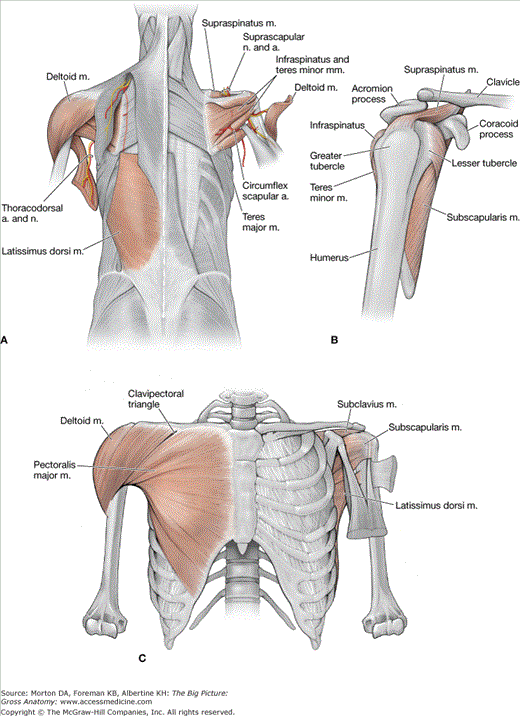
The following muscles and muscle groups comprise the muscles of the glenohumeral joint (Figure 30-3A–C and Table 30–2)
- Deltoid muscle. The deltoid muscle attaches to the spine of the scapula, acromion, clavicle, and the deltoid tuberosity of the humerus. It has a triangular shape, with the muscle fibers coursing anteriorly to the glenohumeral joint producing flexion, laterally producing abduction, and posteriorly producing extension. The deltoid muscle is innervated by the axillary nerve (C5–C6) and receives its blood supply from the thoracoacromial trunk (deltoid and acromial branches), the anterior and posterior humeral circumflex arteries, and the subscapular artery.
- Intertubercular groove muscles. This group of muscles is named because of their common insertion into the intertubercular sulcus of the humerus.
- Pectoralis major muscle. Attaches to the sternum, clavicle, and costal margins and laterally attaches over the long head of the biceps brachii tendon to insert into the lateral lip of the intertubercular groove of the humerus. The pectoralis major muscle is a prime flexor, adductor, and medial rotator of the humerus. The medial and lateral pectoral nerves (ventral rami of C5–T1) provide innervation. The pectoral branch of the thoracoacromial trunk provides most of the blood supply to the pectoralis major muscle.
- Latissimus dorsi muscle. A broad, flat muscle of the lower region of the back. The latissimus dorsi muscle attaches to the spinous processes of T7 inferiorly to the sacrum via the thoracolumbar fascia, and inserts laterally into the intertubercular groove of the humerus. The latissimus dorsi muscle acts on the humerus (arm), causing powerful adduction, extension, and medial rotation of the arm. It is innervated by the thoracodorsal nerve (ventral rami of C6–C8) and receives its blood supply from the thoracodorsal artery (branch off the axillary artery).
- Teres major muscle. Attaches to the inferior angle of the scapula and the medial lip of the intertubercular sulcus. The teres major muscle medially rotates the humerus and is innervated by the lower subscapular nerve (C5–C6).
- Pectoralis major muscle. Attaches to the sternum, clavicle, and costal margins and laterally attaches over the long head of the biceps brachii tendon to insert into the lateral lip of the intertubercular groove of the humerus. The pectoralis major muscle is a prime flexor, adductor, and medial rotator of the humerus. The medial and lateral pectoral nerves (ventral rami of C5–T1) provide innervation. The pectoral branch of the thoracoacromial trunk provides most of the blood supply to the pectoralis major muscle.
- Rotator cuff muscles. The rotator cuff muscles consist of four muscles (supraspinatus, infraspinatus, teres minor, and subscapularis) that form a musculotendinous cuff around the glenohumeral joint. The cuff provides muscular support primarily to the anterior, posterior, and superior aspects of the joint (the first letter of each muscle forms an acronym known as SITS).
- Supraspinatus muscle. Attaches to the supraspinous fossa and courses under the acromion to attach to the greater tubercle of the humerus. Humeral abduction is the primary action of the supraspinatus muscle. The suprascapular nerve (C5–C6) and the suprascapular artery provide innervation and blood supply to the supraspinatus muscle.
- Infraspinatus muscle. Attaches to the infraspinous fossa of the scapula and courses posteriorly to the glenohumeral joint to attach to the greater tubercle of the humerus. The primary action of the infraspinatus muscle is lateral rotation of the humerus. The suprascapular nerve (C5–C6) provides innervation, and the suprascapular artery provides the vascular supply to the infraspinatus muscle.
- Teres minor muscle. Attaches to the lateral border of the scapula and greater tubercle of the humerus. The teres minor muscle courses posteriorly to the glenohumeral joint and produces lateral rotation of the humerus. The axillary nerve (C5–C6) supplies innervation, and the circumflex scapular artery supplies blood to the teres minor muscle.
- Subscapularis muscle. Attaches to the subscapular fossa on the deep side of the scapula. The subscapularis muscle crosses the anterior glenohumeral joint to attach to the lesser tubercle of the humerus, producing medial rotation of the humerus with contraction. The muscle is innervated by the upper and lower subscapular nerves (C5–C6). The suprascapular, axillary, and subscapular arteries supply blood to the subscapularis muscle.
- Supraspinatus muscle. Attaches to the supraspinous fossa and courses under the acromion to attach to the greater tubercle of the humerus. Humeral abduction is the primary action of the supraspinatus muscle. The suprascapular nerve (C5–C6) and the suprascapular artery provide innervation and blood supply to the supraspinatus muscle.
Stay updated, free articles. Join our Telegram channel

Full access? Get Clinical Tree


