Pleura
The lungs exchange oxygen and carbon dioxide and are the functional organs of the respiratory system. To serve that vital function, the lungs are located adjacent to the heart within the pleural sacs. The pleurae are serous membranes that line the internal surface of the thoracic cage and the outside surface of the lungs. The plurae secrete fluid that decreases resistance against lung movment during breathing.
Each lung (right and left) is contained within a serous membrane called the pleural sac (Figure 3-1A). The right and left pleural sacs occupy most of the thoracic cavity and flank both sides of the heart. Each pleural sac is composed of two layers of serous (secretory) membrane, the parietal pleura and the visceral pleura (Figure 3-1B).
- Parietal pleura. The external serous membrane lining the internal surface (wall) of the thoracic cavity.
- Visceral pleura. The internal serous membrane intimately attached to the surface of each lung.
- Pleural fluid. A layer of fluid located between the parietal and visceral pleurae in what is called the pleural space.
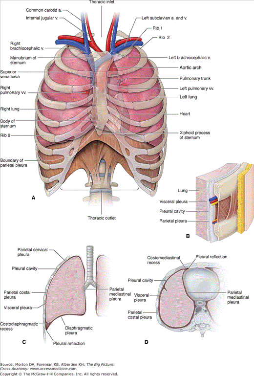
As previously described, the parietal pleura lines the internal surface (wall) of the thoracic cavity, including the lateral sides of the mediastinum (Figure 3-1C and D). The parietal pleura is separated from the thoracic wall by the endothoracic fascia, a thin layer of connective tissue located between the parietal pleura and the innermost intercostal muscles and membrane. The parietal pleura is assigned specific names, depending on the structures that it lines.
- Mediastinal parietal pleura. Lines the lateral surface of the mediastinum (the space between the lungs where the heart is located).
- Costal parietal pleura. Lines the internal surface of the ribs.
- Diaphragmatic parietal pleura. Lines the superior surface of the diaphragm.
- Cervical parietal pleura (cupula). Extends above rib 1 to the root of the neck.
Intercostal nerves supply the costal parietal pleura and the peripheral portion of the diaphragmatic parietal pleura. Phrenic nerves supply the central portion of the diaphragmatic parietal pleura and the mediastinal parietal pleura. The parietal pleura is innervated by general sensory neurons, and therefore, it is sensitive to pain.
The parietal pleura receives its vascular supply via branches of the internal thoracic, superior phrenic, posterior intercostal, and superior intercostal arteries.
The visceral pleura is intimately attached to each lung and follows the contour of the lobes of the lung (Figure 3-1C and D). The visceral pleura is contiguous with the parietal pleura at the site where bronchi, vessels, nerves, and lymphatics pass from the mediastinum into the lung (the root, or hilum, of the lungs). In contrast to the parietal pleura, the visceral pleura is insensitive to pain because its visceral sensory neurons originate from the autonomic vagus nerve (CN X). The visceral pleura receives its blood supply via the bronchial arteries, whereas its venous drainage is via the pulmonary veins.
Each pleural cavity has a pleural space. The pleural space is located between the parietal and visceral pleurae and is a closed space, which means that there is no communication between the right and left pleural spaces. The pleural space contains a thin film of pleural fluid that lubricates the surface of the pleurae and facilitates the movement of the lungs across the thoracic wall and diaphragm during inspiration and expiration.
During expiration, air flows out of the lungs, causing the internal pressure of the lungs to decrease and the lung tissue to potentially collapse. However, the thin layer of pleural fluid, working in conjunction with surfactant in the alveoli, keeps the lungs from collapsing by keeping the visceral pleura coupled to the parietal pleura and the alveoli open, respectively. As a result, the lungs remain inflated, even at the end of a deep expiration.
 If air is introduced into the pleural space as a result of chest trauma (e.g., a knife wound), the coupling between the parietal pleura and visceral pleura may be broken, causing the lung to collapse. This pathology is called a pneumothorax. When blood fills the pleural space, the pathology is called a hemothorax.
In quite respiration, the lung, with its covering of visceral pleura, does not fill the entire pleural sac. The locations where the lung does not completely fill the pleural sac are called pleural recesses. There are two clinically important recesses for the right and left lungs that are termed the same as the reflection next to it (Figure 3-1C and D):
- Costodiaphragmatic recess. The recess where the costal parietal pleura meets the diaphragmatic parietal pleura. This recess is located at the inferior limit of the pleural sac.
- Costomediastinal recess. The recess where the costal parietal pleura meets the mediastinal parietal pleura anteriorly, near the midline.
The pleural recesses are sites where pleural fluid accumulates during quiet breathing. When a deep breath is taken, the greatly expanded lungs push into the recesses, allowing lung volume to increase and, consequently, the pleural fluid becomes displaced around each lung.
Stay updated, free articles. Join our Telegram channel

Full access? Get Clinical Tree


