Bones of the Shoulder and Arm
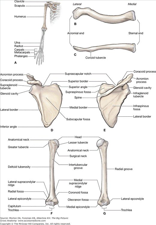
The bones of the skeleton provide a framework to which soft tissues (e.g., muscles) can attach. The bony structure of the shoulder and arm, from proximal to distal, consists of the clavicle, scapula, and humerus (Figure 29-1A). Synovial joints and ligaments connect bone to bone.
The clavicle, or collarbone, is the only bony attachment between the upper limb and the axial skeleton (Figure 29-1B and C). It is superficial along its entire length and shaped like an “S.” The clavicle provides an attachment for muscles that connect the clavicle to the trunk and the upper limb. The following landmarks are found on the clavicle:
- Acromial end. Articulates laterally with the acromion of the scapula and forms the acromioclavicular joint.
- Sternal end. Articulates medially with the manubrium and forms the sternoclavicular joint.
- Conoid tubercle. Located on the inferior surface of the lateral clavicle and serves as an attachment for the coracoclavicular ligament.
The scapula, or shoulder blade, is a large, flat triangular bone with three angles (lateral, superior, and inferior), three borders (superior, lateral, and medial), two surfaces (costal and posterior), and three processes (acromion, spine, and coracoid) (Figure 29-1D and E). The following landmarks are found on the scapula:
- Subscapular fossa. Located anteriorly and characterized by a shallow, concave fossa. Because the subscapular fossa glides upon the ribs, it is also known as the costal surface.
- Acromion. A relatively large projection of the anterolateral surface of the spine; the acromion arches over the glenohumeral joint and articulates with the clavicle.
- Spine. Very prominent and palpable; the spine subdivides the posterior surface of the scapula into a small supraspinous fossa and a larger infraspinous fossa.
- Supraspinous fossa. Located on the posterior surface of the scapula and superior to the spine of the scapula.
- Infraspinous fossa. Located on the posterior surface of the scapula and inferior to the spine of the scapula.
- Suprascapular notch. A small notch medial to the root of the coracoid process where the suprascapular nerve, artery, and vein course.
- Glenoid cavity (fossa). A shallow cavity that articulates with the head of the humerus to form the glenohumeral joint.
- Supraglenoid tubercle. Located superior to the glenoid cavity and serves as the attachment for the long head of the biceps brachii muscle.
- Infraglenoid tubercle. Located inferior to the glenoid cavity and serves as the attachment for the long head of the triceps brachii muscle.
- Coracoid process. A prominent and palpable hook-like structure inferior to the clavicle. The coracoid process serves as an attachment for the pectoralis minor, coracobrachialis, and short head of the biceps brachii muscles.
The humerus is the longest bone of the arm and is characterized by many distinct features that help to allow the upper extremity to move through a significant range of motion. The following landmarks are found on the humerus (Figure 29-1F and G):
- Head. A ball-shaped structure that articulates with the glenoid cavity.
- Anatomical neck. Formed by a narrow constriction immediately distal to the head of the humerus.
- Surgical neck. Lies distal to the anatomical neck and tubercles of the humerus. The axillary nerve and the posterior humeral circumflex artery course into the posterior compartment of the arm, deep to the surgical neck.
- Greater and lesser tubercles. Enlarged areas for muscle attachments.
- Intertubercular (bicipital) groove. A deep sulcus between the greater and lesser tubercles, where the long head of the biceps brachii tendon courses en route to the supraglenoid tubercle.
- Radial (spiral) groove. A distinct groove on the posterior surface of the humerus, where the radial nerve and the deep brachial artery course.
- Deltoid tuberosity. A large V-shaped protrusion on the lateral surface of the humerus, midway along its length where the deltoid muscle attaches.
- Lateral epicondyle. Located on the distal lateral end of the humerus and provides an attachment surface for the posterior forearm muscles (extensors).
- Medial epicondyle. Located on the distal medial end of the humerus and provides an attachment surface for the anterior forearm muscles (flexors).
- Trochlea. Characterized by a pulley shape; it helps to guide the hinge joint. The trochlea of the humerus articulates with the trochlear notch of the ulna.
- Capitulum. Characterized by its oval, convex shape for articulation with the radial head.
- Coronoid fossa. Located on the distal anterior surface of the humerus, where the coronoid process of the ulna articulates.
- Olecranon fossa. Located on the distal posterior surface of the humerus, where the olecranon process of the ulna articulates.
Bones of the Forearm and Hand
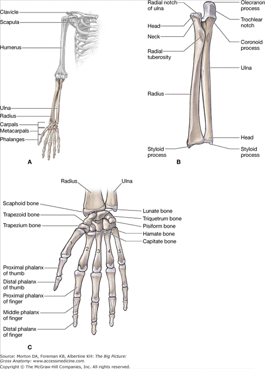
The bony structure of the forearm and hand, from proximal to distal, consists of the radius, ulna, 8 carpals, 5 metacarpals, and 14 phalanges (Figure 29-2A). The radius and ulna are bound together by a tough fibrous sheath known as the interosseous membrane.
In the anatomic position, the radius is the lateral bone of the forearm. It articulates with the capitulum of the humerus and with the ulna. The radius is primarily a bone of movement in the forearm during rotation (supination and pronation) relative to the fixed ulna. The following landmarks are found on the radius (Figure 29-2B):
- Head. A disc-shaped structure that enables the synovial pivot joint in the forearm.
- Radial tuberosity. A swelling inferior to the radial neck on the medial surface that serves as an attachment for the biceps brachii muscle.
- Radial styloid process. Prominent and palpable process on the distal and lateral end that serves as an attachment for the brachioradialis muscle.
In the anatomic position, the ulna is the medial bone of the forearm. It articulates with the trochlea of the humerus and with the radius. The ulna remains relatively fixed during forearm rotation of pronation and supination. The following landmarks are found on the ulna (Figure 29-2B):
- Olecranon process. A large posterior projection that contributes to the trochlear notch and articulates with the humerus (trochlea and the olecranon fossa).
- Coronoid process. A small anterior projection that contributes to the trochlear notch and articulates with the trochlea and the coronoid fossa of the humerus.
- Trochlear notch. A large notch on the proximal end of the ulna that is formed by the olecranon and coronoid processes. It articulates with the trochlea on the humerus.
- Ulnar head. A distal rounded surface at the end of the ulna.
- Ulnar styloid process. A palpable distal projection from the dorsal medial ulna.
Stay updated, free articles. Join our Telegram channel

Full access? Get Clinical Tree


