Overview of the Cranial Nerves
Cranial nerves (CNN) emerge through openings in the skull and are covered by tubular sheaths of connective tissue derived from the cranial meninges. There are 12 pairs of cranial nerves, numbered I to XII, from rostral to caudal, according to their attachment to the brain. The names of the cranial nerves reflect their general distribution and function. Like spinal nerves, cranial nerves are bundles of sensory and motor neurons that conduct impulses from sensory receptors and innervate muscles or glands.
To best understand the cranial nerves, it is helpful to remember the following information:
- Neuron versus nerve. A neuron is a single sensory or motor nerve cell, whereas a nerve is a bundle of neuronal fibers (axons). Cranial nerves have three types of sensory and three types of motor neurons, known as modalities. Therefore, a nerve may be composed of a combination of sensory or motor neurons (e.g., the facial nerve possesses sensory and motor neurons).
- Ganglion. A ganglion is a collection of nerve cell bodies in the peripheral nervous system.
- Nucleus. A nucleus is a collection of nerve cell bodies in the central nervous system (CNS).
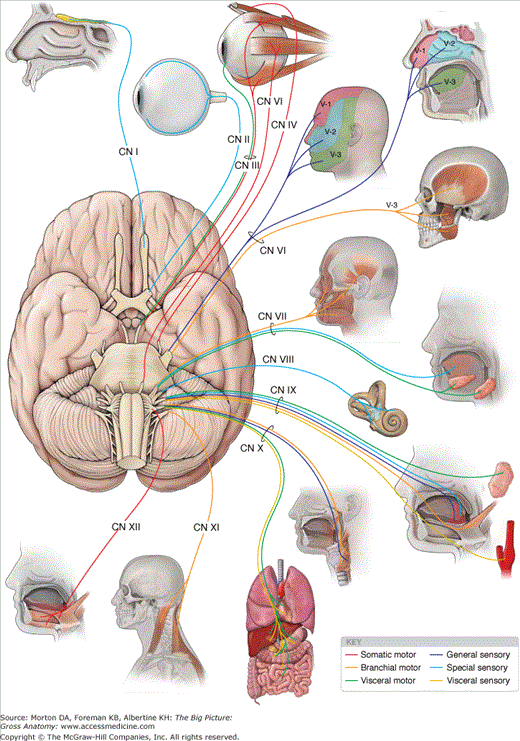
The 12 pairs of cranial nerves may possess one or a combination of the following sensory and motor modalities (Figure 17-1; Table 17-1):
- Sensory (afferent) neurons. Conduct information from the body tissues to the CNS.
- General sensory (general somatic afferent). Transmit sensory information (e.g., touch, pain, and temperature), conducted mainly by CN V but also by CNN VII, IX, and X.
- Special sensory (special visceral afferent). Include special sensory neurons (e.g., smell, vision, taste, hearing, and equilibrium), mainly conducted by the olfactory, optic, and vestibulocochlear nerves (CNN I, II, and VIII, respectively) as well as by CN VII and CN X.
- Visceral sensory (general visceral afferent). Convey sensory information from the viscera, including the gastrointestinal tract, trachea, bronchi, lungs, and heart, as well as the carotid body and sinus. Visceral sensory neurons course within CN IX and CN X.
- General sensory (general somatic afferent). Transmit sensory information (e.g., touch, pain, and temperature), conducted mainly by CN V but also by CNN VII, IX, and X.
- Motor (efferent) neurons. Conduct information from the CNS to body tissues.
- Somatic motor (general somatic efferent) neurons. Innervate skeletal muscles derived from somites, including the extraocular and tongue muscles. Innervation is accomplished via the oculomotor, trochlear, abducens, and hypoglossal nerves (CNN III, IV, VI, and XII, respectively).
- Branchial motor (special visceral efferent) neurons. Innervate skeletal muscles derived from the branchial arches, including the muscles of mastication and facial expression and the palatal, pharyngeal, laryngeal, trapezius and sternocleidomastoid muscles. Innervation is accomplished via the trigeminal, facial, glossopharyngeal, vagus, and spinal accessory nerves (CNN V, VII, IX, X, and XI, respectively).
- Visceral motor (general visceral efferent) neurons. Innervate involuntary (smooth) muscles or glands, including visceral motor neurons that constitute the cranial outflow of the parasympathetic division of the autonomic nervous system. The preganglionic neurons originate in the brainstem and synapse outside the brain in parasympathetic ganglia. The postganglionic neurons innervate smooth muscles and glands via CNN III, VII, IX, and X.
- Somatic motor (general somatic efferent) neurons. Innervate skeletal muscles derived from somites, including the extraocular and tongue muscles. Innervation is accomplished via the oculomotor, trochlear, abducens, and hypoglossal nerves (CNN III, IV, VI, and XII, respectively).
Modality | General Function | CNN Containing the Modality |
|---|---|---|
General sensory | Perception of touch, pain, temperature | CN V (trigeminal), CN VII (facial), CN IX (glossopharyngeal), CN X (vagus) |
Special sensory | Vision, smell, hearing, balance, taste | CN I (olfactory), CN II (optic), CN VII (facial), CN IX (glossopharyngeal) |
Visceral sensory | Sensory input from viscera | CN IX (glossopharyngeal), CN X (vagus) |
Branchial motor | Motor innervation to skeletal muscle derived from branchial arches | CN V-3 (mandibular), CN VII (facial), CN IX (glossopharyngeal), CN X (vagus), CN XI (spinal accessory) |
Somatic motor | Motor innervation of skeletal muscle derived from somites | CN III (oculomotor), CN IV (trochlear), CN VI (abducens), CN XII (hypoglossal) |
Visceral motor | Motor innervation to smooth muscle, heart muscle, and glands | CN III (oculomotor), CN VII (facial), CN IX (glossopharyngeal), CN X (vagus) |
The nuclei of the cranial nerves (where motor neurons originate or sensory neurons terminate) are located in the brainstem, with the exception of CN I and CN II, which are extensions of the forebrain.
The specific functions of cranial nerves depend on the nature of the anatomic targets of the cranial nerves (Table 17-2).
- CNN V, VII, IX, and X. Innervate almost all of the structures of the head and neck, such as the skin, mucous membranes, muscle, and glands derived from the pharyngeal arches. Of these four nerves, CN V and CN VII innervate most of these structures, whereas CN IX innervates only a few structures of the head, oral cavity, pharynx, and neck. Almost all targets of CN X are in the trunk.
- CNN III, IV, and VI. Innervate only structures in the orbit.
- CNN I, II, and VIII. Possess only special sensory neurons for smell, sight, balance, and hearing.
- CN XII. Innervates only the tongue muscles.
- CNN III, VII, IX, and X. The cranial nerves that carry parasympathetic neurons.
CN | Modalities and Function | Exit from Skull |
|---|---|---|
CN I (olfactory) | Special sensory: smell | Cribriform plate of the ethmoid bone |
CN II (optic) | Special sensory: sight | Optic canal |
CN III (oculomotor) | Somatic motor: levator palpebrae superioris m.; superior, medial, and inferior rectus mm.; inferior oblique mm. Visceral motor: sphincter pupillae m. (pupil constriction) and ciliary mm. (lens accommodation) | Superior orbital fissure |
CN IV (trochlear) | Somatic motor: superior oblique m. | Superior orbital fissure |
CN V (trigeminal) | General sensory: CN V-1: orbit and forehead CN V-2: maxillary region CN V-3: mandibular region, tongue Branchial motor: CN V-3: muscles of mastication, mylohyoid, anterior digastricus, tensor tympani, and tensor veli palatine mm. | CN V-1: superior orbital fissure CN V-2: foramen rotundum CN V-3: foramen ovale |
CN VI (abducens) | Somatic motor: lateral rectus m. | Superior orbital fissure |
CN VII (facial) | General sensory: external acoustic meatus and auricle Special sensory: anterior two-thirds of tongue Branchial motor: muscles of facial expression and stylohyoid, posterior digastricus, stapedius mm. Visceral motor: all glands of the head (lacrimal, submandibular, sublingual, palatal, nasal) except the one it courses through (does not innervate the parotid) | Internal acoustic meatus |
CN VIII (vestibulocochlear) | Special sensory: hearing, balance, and equilibrium | Internal acoustic meatus |
CN IX (glossopharyngeal) | General sensory: posterior third of tongue, oropharynx, tympanic membrane, middle ear, and auditory tube Special sensory: taste from posterior one-third of tongue Visceral sensory: carotid sinus (baroreceptor) and carotid body (chemoreceptor) Branchial motor: stylopharyngeus m. Visceral motor: parotid gland | Jugular foramen |
CN X (vagus) | General sensory: skin of the posterior ear and external acoustic meatus Visceral sensory: aortic and carotid bodies (chemoreceptors) and aortic arch (baroreceptor) Branchial motor: all palatal muscles (except tensor tympani); all pharyngeal muscles (except stylopharyngeus m.) and all laryngeal mm. Visceral motor: heart, smooth muscle, and glands of the respiratory tract, gastrointestinal tube, and viscera of the foregut and midgut | Jugular foramen |
CN XI (spinal accessory) | Branchial motor: trapezius and sternocleidomastoid mm. | Jugular foramen |
CN XII (hypoglossal) | Somatic motor: tongue mm. (except palatoglossus m.) | Hypoglossal canal |
CN I: Olfactory Nerve
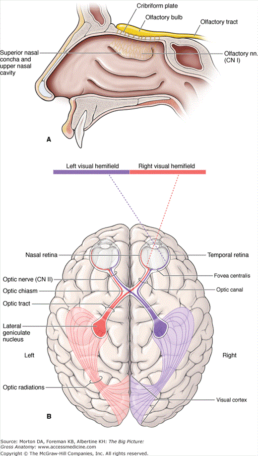
The olfactory neurons originate in the olfactory epithelium in the superior part of the lateral and septal walls of the nasal cavity. The nerves ascend through the cribriform foramina of the ethmoid bone to reach the olfactory bulbs. The olfactory neurons synapse with neurons in the bulbs, which course to the primary and association areas of the cerebral cortex.
 Injury to CN I (e.g., a fracture of the cranial base) can result in anosmia (loss of smell), tearing of the meninges, or cerebrospinal fluid rhinorrhea.
CN II: Optic Nerve
Optic nerve fibers arise from the retina and all converge at the optic disc. CN II exits the orbit via the optic canals. Both optic nerves form the optic chiasm, the site where neurons from the nasal side of either retina cross over to the contralateral side of the brain. The neurons then pass via the optic tracts to the thalamus, where they synapse with neurons that course to the primary visual cortex of the occipital lobe.
- Visual fields. The visual field is the part of the world seen by the eyes. The entire visual field is divided into right and left and upper and lower regions, defined when the patient is looking straight ahead. Each eye has its own visual field or, in other words, the part of the world seen by each eye alone. The lateral visual field of an eye is called the temporal field, whereas the medial visual field of the same eye is called the nasal field. Although the fields of vision of the two eyes overlap greatly, the right eye sees things far to the right that the left eye cannot see, and vice versa.
- Optic chiasm. Each optic nerve carries axons from the entire retina of the eye. However, after coursing through their respective optic canals, the right and left optic nerves engage in a redistribution of axons at the optic chiasma, located just anterior to the pituitary stalk. The optic chiasma is created by neurons from the nasal half of each retina crossing over to the opposite side.
- Optic tracts. The two optic tracts emerge from the optic chiasma. The right optic tract contains axons from the temporal half of the right retina and the nasal half of the left retina and carries information about the entire left visual field. The left optic tract contains neurons from the temporal half of the left retina and the nasal half of the right retina and carries information about the entire right visual field. An optic tract is named according to the side of the body on which it lies, but it is concerned with the contralateral visual field.
 Injury to CN II may result in monocular blindness. If an optic tract is injured, the result is hemianopia; that is, half the visual field of each eye is lost. Specifically, the half-field of one eye that is lost is the same side as the half-field that is lost by the other eye. The lost fields will be contralateral to the damaged optic tract. For example, interruption of the function in the left optic tract causes loss of vision in the right visual fields of both eyes. In addition, injury to CN II may result in a loss of the papillary reflex of CN III.
 Most cranial nerves are peripheral nerves and therefore myelinated by Schwann cells. However, CN II is an extension of the forebrain and as such is myelinated by oligodendrocytes. Multiple sclerosis is an autoimmune disorder that attacks myelin in oligodendrocytes. Therefore, CN II is the only cranial nerve affected by multiple sclerosis.
CN III: Oculomotor Nerve
The oculomotor nerve innervates the levator palpebrae superioris muscle, four of the six extraocular muscles as well as the pupillary constrictor and ciliary muscles.
Upon exiting the midbrain, the oculomotor nerve courses between the posterior cerebral and superior cerebellar arteries and runs along the lateral wall of the cavernous sinus superior to CN IV (Figure 17-3A). CN III enters the orbit through the superior orbital fissure, where it divides into superior and inferior divisions. CN III has the following modalities:
- Somatic motor neurons. CN III innervates four of the extraocular muscles (superior rectus, medial rectus, inferior rectus, and inferior oblique) and the levator palpebrae superioris muscle. The somatic motor component of CN III plays a major role in controlling the muscles responsible for the precise movements of the eyes.
- Visceral motor neurons. Preganglionic parasympathetic neurons of CN III originate in the Edinger–Westphal nucleus and synapse in the ciliary ganglion, providing innervation to the ciliary body (lens accommodation) and sphincter pupillae muscle (pupil constriction) (Figure 17-3B).
Stay updated, free articles. Join our Telegram channel

Full access? Get Clinical Tree


