Anatomy of the Scalp
The scalp consists of five layers of tissue. The five layers, from superficial to deep, are skin, subcutaneous connective tissue, a muscular aponeurotic layer, a loose connective tissue layer, and the pericranium.
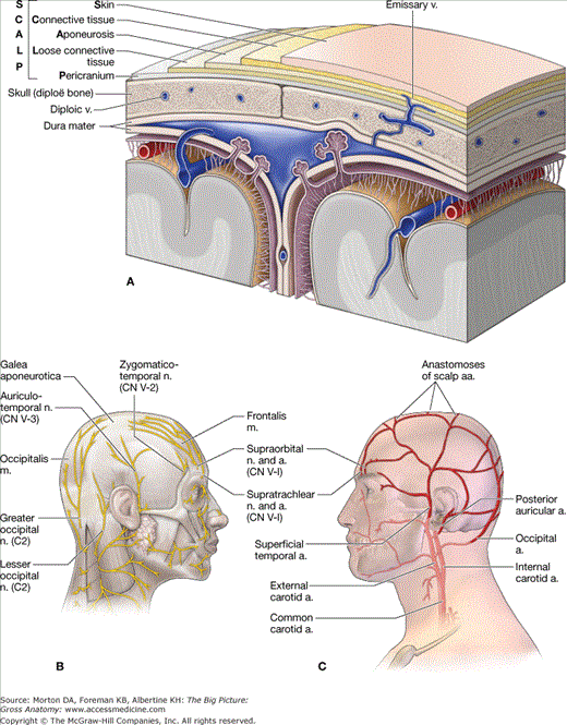
The layers of the scalp can best be remembered by the acronym “SCALP,” with each letter of the word representing the tissue layer associated with it Figure 15-1A.
- Skin. The skin of the scalp contains sweat and sebaceous glands and usually numerous hair follicles.
- Connective tissue. The tissue between the skin and the aponeurotic layers is composed of dense collagenous connective tissue and contains the arteries, veins and nerves supplying the scalp.
- Aponeurosis. The superficial musculoaponeurotic system of the scalp consists of the occipitofrontalis muscle and its investing fascia. This fascia is specialized to form a tendinous epicranial aponeurosis known as the galea aponeurotica. The galea continues into the temples, investing the auricular muscles, and terminates by attaching to the mastoid processes and the zygomatic arch. The frontalis muscle is instrumental in movements of the eyebrows and forehead and is an important muscle of facial expression are innervated by the facial nerve, cranial nerve (CN) VII.
- Loose connective tissue. A sponge-like layer of loose connective tissue forms a subaponeurotic compartment that enables free movement of the top three scalp layers across the pericranium. It also contains the emissary veins.
- Pericranium. The pericranium is the periosteum over the external surface of the skull where the fibrous tissue knits into the sutures.
 Understanding the structure of the scalp is important when treating patients with scalp wounds. Superficial scalp wounds do not gape because of the strength of the underlying aponeurosis, which holds the margins of the wound together. However, if the aponeurosis is lacerated in the coronal plane, deep scalp wounds gape because of the contraction of the frontalis and occipitalis muscles, which contract in opposite directions. This aponeurotic layer of the scalp is often tightened during cosmetic surgery (e.g., “facelifts”) to help reduce wrinkles in the face and forehead.
 Injury to the fourth layer of the scalp (loose connective tissue) is dangerous because infection can potentially spread from the scalp through emissary veins into the cranial cavity. In addition, an infection or fluid can enter the eyelids because the frontalis muscle inserts into the skin and subcutaneous tissue (not to bone), resulting in ecchymosis, or “black-eyes.”
The scalp receives its cutaneous innervation as follows (Figure 15-1B):
- Posterior region of the scalp. Innervated by branches of the cervical plexus, which are principally derived from C2 and C3 (lesser occipital and greater occipital nerves).
- Anterior region of the scalp. Innervated by the supraorbital and supratrochlear nerves, which are derived from the ophthalmic division of the trigeminal nerve (CN V-1).
- Lateral region of the scalp. Innervated by branches of the maxillary (CN V-2) and mandibular (CN V-3) divisions of the trigeminal nerve (the zygomaticotemporal and auriculotemporal nerves, respectively).
The scalp is highly vascularized via branches of the external and internal carotid arteries (Figure 15-1C).
- External carotid artery. Branches include the occipital, posterior auricular, and superficial temporal arteries.
- Internal carotid artery. Branches include the supraorbital and supratrochlear arteries.
 Scalp lacerations usually bleed profusely, primarily because arteries enter the scalp and bleed from both ends of the artery as a result of abundant anastamoses. In addition, the severed arteries do not contract when they are cut because the vessel lumens are held open by the dense connective tissue in the second layer of the scalp. As a result, bleeding from the scalp can be profuse.
The supraorbital and supratrochlear veins unite to form the facial vein. The superficial temporal vein joins with the maxillary vein to form the retromandibular vein in the parotid salivary gland. The posterior auricular vein unites with the posterior division of the retromandibular vein to form the external jugular vein.
Scalp veins connect with the diploic and emissary veins.
- Diploic veins. Diploic veins course in the diploe cranial bones of the skull and connect with the dural venous sinuses via emissary veins.
- Emissary veins. Small veins connect veins of the scalp and skull with the dural venous sinuses.
Skull
There are 8 cranial bones and 14 facial bones in the skull, all of which serve to protect the brain.
The skull protects the brain and its surrounding meninges (Figure 15-2A). The outer and inner surfaces of the skull, are covered by periosteum, known respectively as the pericranium and the endocranium. The periosteum is continuous at the sutures of the skull. The cranial bones consist of spongy bone “sandwiched” between two layers of compact bone. The bones of the skull are as follows (Figure 15-2B–E):
- Frontal bone. The unpaired frontal bone underlies the forehead, roof of the orbit, and a smooth median prominence called the glabella.
- Parietal bone. The paired parietal bones form the superior and lateral aspects of the skull.
- Temporal bone. The paired temporal bones consist of a squamous part that forms the lateral portion of the skull; the petrous part, which encloses the internal ear (cochlea and semicircular canals) and the middle ear (malleus, incus, and stapes); the mastoid part, which contains the mastoid air cells; and the tympanic part, which houses the external auditory meatus and tympanic cavity.
- Occipital bone.

Stay updated, free articles. Join our Telegram channel

Full access? Get Clinical Tree


