Inclusion criteria
Age between 8 and 17 years
Children with diagnosed asthma, confirmed by spirometry
Children treated with inhaled corticosteroids continuously for at least 4 years
The time without treatment was never longer than 3 months
Signing a consent form
Exclusion criteria
Children < 8 and > 17 years old
Children with severe health problems, such as mental illness (schizophrenia, depression, post-traumatic stress, bipolar disorder, addiction), disease with neurological symptoms (seizures), disease causing chronic hypoxia (congenital heart defects), prematurity <37 Hbd and Apgar score ≤6 points at 1 and 5 min
Contraindication to MRI (metallic implants, claustrophobia)
Refusal to sign a consent form
The MR images were performed on a 1.5 T scanner with parallel imaging capability (12 channels, Toshiba, Japan). Structural images consisted of axial T1 (repetition time-TR 650 ms/echo time-TE 17 ms), T2 (TR 5,200 ms/TE 105 ms), using a turbo spin echo (5 mm slices with 1 mm interslice gap), FLAIR (TR 8,000 ms/TE 105 ms), and DWI sequences using typical parameters. DWI were sensitized in six collinear directions at an effective b value of 1,000 s/mm2. Images were assessed in the three planes: axial, coronal, and sagittal. All examinations were evaluated by two independent readers: a neuroradiologist and a pediatrician.
3 Results
3.1 Study Group
All patients, aside from asthma, suffered from allergic rhinitis, and four of them had atopic dermatitis. One child was under the care of a nephrologist due to IgA nephropathy. Six children, in addition to ICS, was treated with intranasal steroids (intermittent or persistent), and another five with allergen immunotherapy. Steroid therapy was continuing from 4 to 10 years, with a medium dose in six patients and a low dose in three patients as based on GINA recommendations (2008). Physical and neurological examinations were normal in all patients. At the time of the study, asthma was under control in all of the patients (Asthma Control Test 24-25 points) and spirometry was normal.
3.2 Magnetic Resonance Imaging
The MRI images showing no cortical abnormalities in asthmatic children treated with ICS are presented in Fig. 1 to be compared with the pathological features found in some children. In six patients, the MRI studies revealed small subcortical hyperintense foci. Three of these patients had more than five lesions, all of which were smaller than 3 mm. Examples of such lesions are shown in Fig. 2a and b. Features of mild supratentorial cortical atrophy were apparent in four patients, as exemplified in Fig. 3. The cerebellum was unremarkable in all the children imaged.
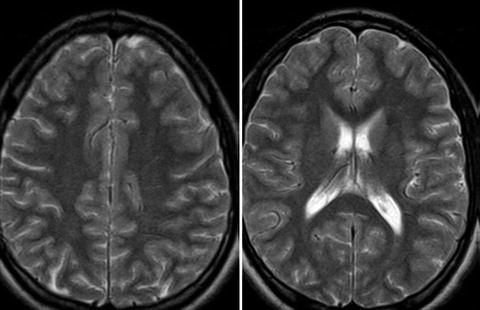
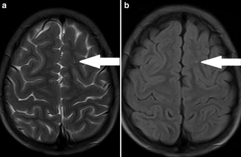
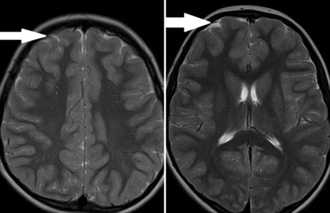
Fig. 1
T2 weighted axial images. Normal brain of a 11 years old boy. No atrophy or white matter abnormalities are noted
Fig. 2
Small lesion with increased signal intensity in T2 weighted (a) and FLAIR (b) images, located in the subcortical white matter of the frontal lobe (arrows)
Fig. 3


Mild cortical atrophy of frontal lobes in otherwise normal brain of a 9 years old child. T2 weighted axial images (arrows)
Stay updated, free articles. Join our Telegram channel
Full access? Get Clinical Tree
