Cellular Growth Control and Cancer

Chapter 18 Cellular Growth Control and Cancer


The human body is produced from the fertilized ovum in a succession of mitotic cell divisions. Each mitotic cycle consists of an orderly sequence of events, including growth, DNA replication, and cell division.


As they go through repeated rounds of cell division, the totipotent cells of the embryo metamorphose into the differentiated cells of the mature body: blood cells, neurons, muscle cells, and so forth. Cell growth, mitotic rate, and cell differentiation are controlled by external stimuli, including nutrients, hormones, growth factors, and contacts with neighboring cells and the extracellular matrix.


Derangements in the controls on the cell’s proliferation, differentiation, and survival cause cancer. This chapter describes the elements of cell cycle control and their abnormalities in cancer.


The cell cycle is controlled at two checkpoints


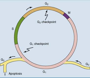
Under the microscope, only two phases of the cell cycle can be distinguished in dividing cells: interphase and mitosis (Fig. 18.1). Mitosis, which lasts between 1 and 4 hours, is the stage of cell division. All of the rest is interphase. Chromosomes are visible as distinct entities only during mitosis, when the DNA is packaged for relocation into the daughter cells. During interphase, there is only dispersed chromatin all over the nucleus.


Interphase is subdivided into three phases. G1 phase (G for gap) is the regular, diploid state of the cell. G1 is followed by S phase (S for synthesis), during which the DNA is replicated, and finally by G2. S phase can be identified by feeding the cells with radiolabeled thymidine. Cells in S phase, which lasts about 6 hours, incorporate a large amount of the thymidine into DNA (but not RNA). Outside S phase, only a small amount of DNA synthesis takes place during DNA repair.


The cell makes three important all-or-none decisions during the cell cycle. At the G1 checkpoint in late G1, it decides about entry into S phase. DNA replication should be initiated only when the cell is ready to progress through the complete cell cycle and only after any DNA damage that may have been sustained has been thoroughly repaired. The overall rate of cell proliferation is controlled by G1.


At the G2 checkpoint in late G2, the cell decides about entry into mitosis. Mitosis should begin only after the completion of DNA replication and only if the replicated chromosomes are structurally intact.


The spindle assembly checkpoint, finally, ensures that the cell proceeds from mitotic metaphase to anaphase only if the mitotic spindle is intact and all chromosomes are attached to the spindle fibers.


Nondividing cells are said to be in G0. Some nondividing cells, including neurons and skeletal muscle fibers, are in G0 forever. Others, including fibroblasts, hepatocytes, and lymphocytes, are usually in G0 but can be coaxed into the cell cycle by growth factors or, in the case of lymphocytes, by antigen along with cytokines from helper T cells.


Cyclins play key roles in cell cycle control


Cell cycle progression depends on the phosphorylation of multiple proteins by the cyclin-dependent kinases (Cdks) in the nucleus and their regulatory subunits, the cyclins. The Cdks are present more or less at all times, but most of the cyclins come and go with the phases of the cell cycle (Fig. 18.2).
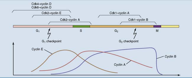


Only cyclin D is not controlled by the cell cycle; it is controlled by mitogens. It rises when a cell in G1 is exposed to mitogens. By activating Cdk4 and Cdk6, cyclin D induces the synthesis of cyclin E, an activator of Cdk2. Cyclin E and Cdk2 push the cell through the G1 checkpoint. Next comes cyclin A, which activates Cdk1 (formerly called Cdc2) and Cdk2. It brings the cell through S phase and remains active through G2.


Finally cyclin B, working with Cdk1, accumulates during G2 and early mitosis. It condenses the chromosomes by phosphorylating chromosomal scaffold proteins and histone H1, and it breaks down the nuclear envelope by phosphorylating and thereby dismantling the lamin network under the inner nuclear membrane. At the spindle assembly checkpoint in mitosis, a ubiquitin ligase complex is formed that destroys cyclin B along with cyclin A and some other proteins of early mitosis suddenly during the metaphase-anaphase transition.


The Cdks are controlled primarily by cyclins, but also by stimulatory and inhibitory phosphorylations and by Cdk inhibitors that are formed under the influence of antimitotic agents.


Retinoblastoma protein guards the G1 checkpoint


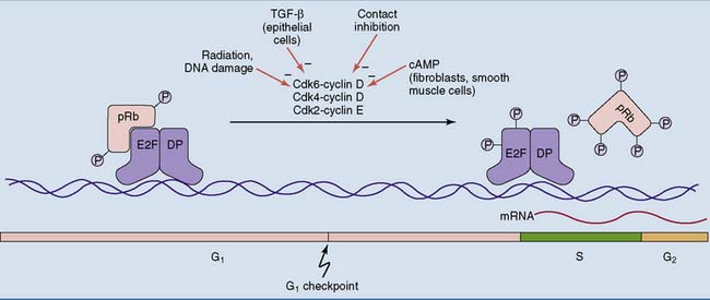
Progression through the cell cycle and DNA synthesis require the transcription factor E2F, which regulates more than 500 genes. It activates the transcription of genes for cyclins D1 (there are three closely related D cyclins), E, A, and B, Cdk1, thymidylate synthetase, dihydrofolate reductase, DNA polymerase α, topoisomerase II, the clamp protein PCNA (see Chapter 7), and the proto-oncogenes MYC and MYCL1 (which code for the transcription factors c-Myc and N-Myc, respectively).


In quiescent cells, E2F is prevented from activating the transcription of these genes by the retinoblastoma protein (pRb), which is encoded by the RB1 gene (Fig. 18.3). Throughout G0 and early G1, pRb is tightly bound to E2F. It prevents gene expression by masking the transcriptional activation domain of E2F and by recruiting a histone deacetylase. At the G1 checkpoint, however, both pRb and E2F become phosphorylated by the kinase complexes of cyclins D and E. The phosphorylated pRb falls off the transcription factor, and the genes can be transcribed.


These events are all or none because they are subject to positive feedback. Once the activity of the cyclin D-Cdk complexes has passed a threshold, the cyclin genes become derepressed. Even more cyclin-Cdk is formed, pushing the cell through the G1 checkpoint.


Cell proliferation is triggered by mitogens


During embryonic development, cell proliferation is tightly linked to cell differentiation. Undifferentiated stem cells divide frequently, but once the cell has morphed into a specialized cell type, it withdraws from the cell cycle. For some cells, including neurons and skeletal muscle fibers, the withdrawal into G0 is final. However, other cells, including hepatocytes and fibroblasts, behave like Sleeping Beauty. They can be restored to reproductive life by external agents. The prince’s kiss that causes these cells to abandon G0 and reenter the cell cycle is delivered by agents called mitogens.


Mitogenic stimuli can be provided by the extracellular matrix. The integrins in focal adhesions (see Chapter 13) not only mediate adhesion to extracellular matrix components but also provide an assembly point for signaling molecules. Cell-matrix contacts tend to be mitogenic, but cell-cell contacts usually are antimitogenic.


Soluble growth factors allow the cell to respond to signals from more distant sources. The term is used loosely to refer to proteins that stimulate cell growth (growth factors in the strict sense), cell proliferation (mitogens), or cell survival (survival factors). Examples of growth factors include the following:


5. Erythropoietin is released from the kidney in response to hypoxia. Acting on a JAK-STAT coupled receptor (see Chapter 17), it stimulates specifically the development of red blood cell precursors in the bone marrow.


Mitogens regulate gene expression


Mitogens push cells through the G1 checkpoint. Figure 18.4 shows two mitogenic signaling cascades that are triggered by autophosphorylated growth factor receptors and activate the nuclear cyclin D-Cdk complexes.


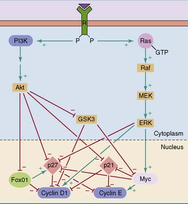
One of these cascades signals through phosphoinositide 3kinase (PI3K) and protein kinase B (PKB, or Akt) (see Chapter 17). PKB phosphorylates and thereby inhibits another protein kinase, glycogen synthase kinase 3 (GSK3). GSK3 inhibits the expression of cyclin D1 by phosphorylating transcriptional regulators bound to the promoter of the cyclin D1 gene. By inhibiting these inhibitory phosphorylations, PKB stimulates the expression of the cyclin D1 gene.


The other mitogenic cascade shown in Figure 18.4 is the mitogen-activated protein (MAP) kinase cascade. It starts with activation of the small G protein Ras at the cytoplasmic surface of the plasma membrane (see Chapter 17). Three isoforms of Ras occur in human tissues.


Activated Ras recruits the serine-threonine protein kinase Raf to the plasma membrane, where it becomes activated by phosphorylation. Some isoenzymes of protein kinase C can activate Ras-bound Raf.


Raf phosphorylates and thereby activates the protein kinases MEK1 and MEK2 (MAPK/ERK kinases). The MEKs phosphorylate the serine-threonine kinases ERK1 and ERK2 (extracellular signal-regulated kinases) on threonine and tyrosine residues in the sequence Thr-Glu-Tyr. The ERKs are also known as MAP kinases.


The activated MAP kinases phosphorylate proteins in both the cytoplasm and the nucleus. They regulate transcription factors by phosphorylation, both directly and indirectly by phosphorylating nuclear protein kinases. The products of some of the activated genes, including the proto-oncogene MYC, are themselves regulators of transcription. In addition to the cyclins, cyclin-Cdk inhibitors including p21 and p27 are regulated both by phosphorylation and at the transcriptional level.


Negative controls on mitogenic signaling include the dephosphorylation of proteins by protein phosphatases at all levels, from the autophosphorylated growth factor receptors to the phosphorylated transcription factors. Another negative control is the hydrolysis of its bound GTP by the Ras protein. The GTPase activity of Ras is stimulated by regulatory proteins, including neurofibromin (Clinical Example 18.2).


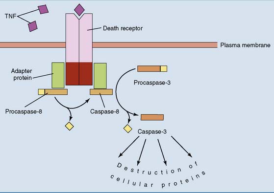
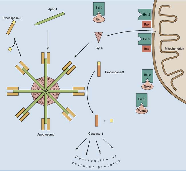
Cells can commit suicide


When a cell dies by necrosis, the environment gets polluted with proteases and other damaging and inflammation-inducing proteins that leak out of the dying cells. Programmed cell death by apoptosis, by contrast, is a clean process in which the dying cell presents itself to macrophages with its membrane intact.


Apoptosis is a normal part of early human development. In adults it remains important as a response to cellular damage, viral infections, somatic mutations, hormonal influences, or lack of extracellular survival factors. Apoptosis eliminates many virus-infected and genetically altered cells. These cells must be prevented from evolving into cancer cells.


Apoptotic stimuli destroy the cell by recruiting proteases of the caspase family. Caspases are present in the cell as inactive precursors (procaspases) that have to be activated by a proteolytic cascade.


Initiator procaspases are activated by death-promoting stimuli. Once activated, their main function is the activation of executioner procaspases that destroy target proteins in the cell. Cleavage of nuclear lamins destroys the nuclear envelope; degradation of a DNase inhibitor unleashes a DNase that cleaves DNA in the spacers between nucleosomes; and cleavage of cytoskeletal and cell adhesion proteins causes the cell to curl up and detach from neighboring cells. This facilitates the removal of the dying cell by macrophages. There are two apoptotic pathways:


1. The extrinsic pathway is triggered by external agents that activate death receptors on the cell surface (Fig. 18.5). These receptors contain a death domain in their cytoplasmic portion. Once activated by an extracellular signal, for example, tumor necrosis factor (TNF) or the Fas ligand, the death domain binds the precursor of an initiator caspase (either Casp-8 or Casp-10) through adapter proteins. Binding to this complex activates the bound procaspases allosterically, enabling them to cleave each other into the active caspases. The activated initiator caspases proceed to activate Casp-3 and other executioner caspases.


Cancers are monoclonal in origin


Because they are genetically identical and depend on one another for transmission of their genes into the next generation, the cells of the human body behave unselfishly toward one another. Each cell grows and divides only to the extent that it furthers the greater good of the body, and some cells even die dutifully—by apoptosis—once their task has been fulfilled.


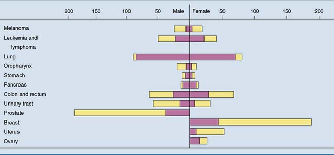
Cancer cells, however, have the cellular equivalent of antisocial personality disorder. A cancer cell arises when a somatic mutation creates a “selfish gene” that causes the cell to proliferate without regard for the greater good of the organism. This single abnormal cell grows into a cell mass called a neoplasm or tumor. Most tumors are monoclonal in origin. This means that all tumor cells are derived from a single abnormal ancestor. Benign tumors limit their growth without doing much harm, but malignant tumors, commonly called cancer, kill the organism. Cancer causes more than 20% of all deaths in industrialized countries. Figure 18.7 shows the incidence of various kinds of cancer in the United States.


Malignant cells retain morphological and biochemical features typical for their cells of origin. Some tumors of epithelial origin, for example, known as carcinomas, still produce keratins; some connective tissue tumors, known as sarcomas, still produce constituents of the extracellular matrix; and some endocrine tumors still secrete hormones. However, these specialized features tend to get lost when cancers become more malignant. Characteristic differences between cancer cells and the normal cells from which they are derived include the following:


Cancer is caused by activation of growth-promoting genes and inactivation of growth-inhibiting genes


Some gene products, including growth factors, growth factor receptors, components of mitogenic signaling cascades, and the G1 cyclins, promote cell proliferation. Others, including cyclin-Cdk inhibitors and pRb, are inhibitory. Therefore two types of mutation can favor mitosis (Fig. 18.8):
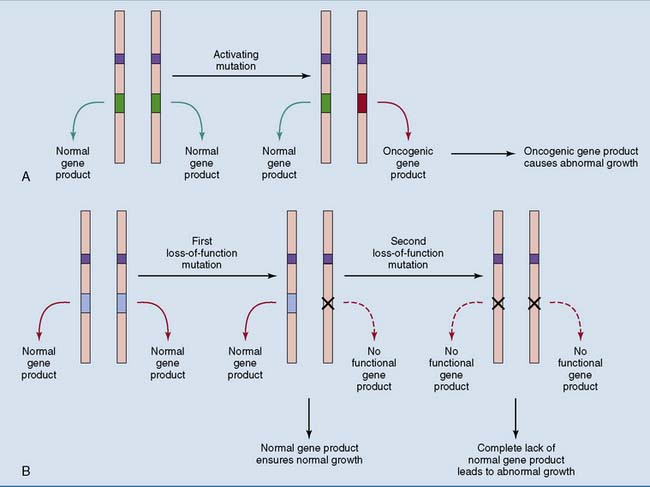


A single mutation is rarely sufficient to convert a cell to malignancy. Common cancers contain a whole assortment of activated oncogenes and inactivated tumor suppressor genes.


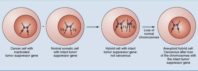
Inactivation of tumor suppressor genes is even more important than activation of cellular proto-oncogenes in most spontaneous cancers. This can be demonstrated in cultured cells (Fig. 18.10). When a cancer cell is fused with a normal cell, the resulting hybrid cell grows like a normal cell because the intact tumor suppressor gene from the normal cell produces the tumor-suppressing protein. If malignant transformation were caused by dominantly acting oncogenes, the hybrid cells would grow like cancer cells.


Jun 18, 2016 | Posted by in BIOCHEMISTRY | Comments Off on Cellular Growth Control and Cancer

Full access? Get Clinical Tree

Get Clinical Tree app for offline access