Cavernous Hemangioma
Brock Kaya, MD
Key Facts
Terminology
Angiographically occult vascular malformation composed of uniform blood vessels with fibrous walls
Clinical Issues
Account for 10-15% of all vascular malformations
Majority (85%) are supratentorial
In children, most common manifestation is hemorrhage
Image Findings
“Popcorn ball” appearance with mixed hyperintense and hypointense signal intensity on both T1- and T2-weighted sequences
Typical low signal intensity ring surrounding lesion due to hemosiderin
Macroscopic Features
Usually small, red-purple, lobulated, mulberry-like lesions
Range in size: Millimeters to several centimeters (diameter)
Microscopic Pathology
Circumscribed, compact aggregate of dilated, thick-walled blood vessels with hyalinized walls
Vascular walls lack smooth muscle and elastic lamina
Little to no intervening brain parenchyma present between blood vessels
Hemosiderin deposition and gliosis in surrounding brain parenchyma
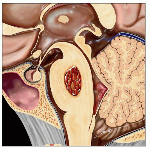 Graphic depicts a cavernous hemangioma of the pons, consisting of a compact aggregate of dilated blood vessels with surrounding hemosiderin deposits. |
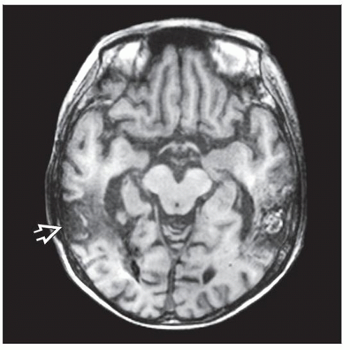 Axial T1WI MR demonstrates a left temporal cavernous hemangioma with central bright T1 contents. A cavernoma  was previously resected on the right. (Courtesy S. M. Holmes, MD.) was previously resected on the right. (Courtesy S. M. Holmes, MD.) |
TERMINOLOGY
Synonyms
Cavernous angioma, cavernous malformation, cavernoma
Definitions
Angiographically occult vascular malformation composed of uniform blood vessels with fibrous walls
ETIOLOGY/PATHOGENESIS
Acquired Anomaly
Previously considered congenital, but now favored to be acquired
Familial forms
Autosomal dominant with incomplete penetrance
Related to mutations in either KRIT1 (CCM1), CCM2, or PDCD10 (CCM3)
May occur secondarily following radiation therapy for childhood brain tumors
CLINICAL ISSUES
Epidemiology
Incidence: 0.5-0.7%
Account for 10-15% of all vascular malformations
Equal sex distribution
Between 9-25% of cases occur in children
Sporadic and familial forms
Sporadic lesions typically solitary
Multiple cavernomas either familial or sporadic
May have associated venous angioma (developmental venous anomaly)
Site
Found throughout CNS
Majority (85%) are supratentorial
Subcortical white matter is most common site
Pons and cerebellum are common infratentorial sites
Presentation
Increasingly, an incidental finding by imaging
Most are asymptomatic
Symptomatic individuals (25% of those affected) may present at any age but most common in 2nd to 5th decades
Seizures
Focal neurologic deficits
Hemorrhage
In adults, uncommon cause of clinically significant cerebral hemorrhage (risk of 0.25-6% per year)
In children, most common manifestation is hemorrhage
Treatment
Observation, surgery, or radiosurgery
In children, surgical excision usually performed due to increased risk for macrohemorrhage
Stay updated, free articles. Join our Telegram channel

Full access? Get Clinical Tree


