Carcinoid Tumor, Colon
Scott R. Owens, MD
Key Facts
Clinical Issues
More commonly occurs in rectum than in remainder of colon
Most are asymptomatic
Local excision sufficient for small tumors
All carcinoids potentially malignant
Microscopic Pathology
Monotonous cells arranged in nests, cords, ribbons
Angiolymphatic invasion common, even in well-differentiated tumors
Ancillary Tests
Neuroendocrine lineage confirmed with synaptophysin and chromogranin immunostains
Top Differential Diagnoses
Adenocarcinoma
Lymphoma
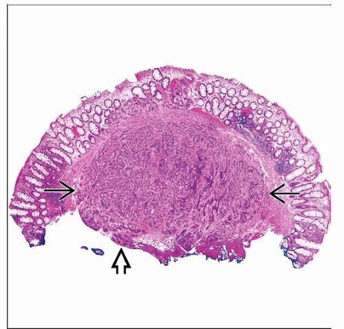 Hematoxylin & eosin shows a carcinoid tumor  centered in the submucosa of a “polyp” that was biopsied during a routine endoscopy. The tumor is present at the cauterized edge centered in the submucosa of a “polyp” that was biopsied during a routine endoscopy. The tumor is present at the cauterized edge  of the biopsy specimen. of the biopsy specimen. |
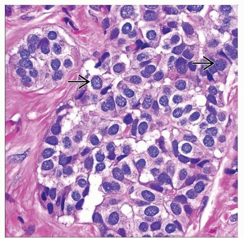 Hematoxylin & eosin shows a high-power view of tumor nests in a carcinoid tumor. Note characteristic “salt and pepper” chromatin distribution  and the homogeneous appearance of the cells. and the homogeneous appearance of the cells. |
TERMINOLOGY
Synonyms
Well-differentiated neuroendocrine neoplasm
Well-differentiated neuroendocrine carcinoma (“malignant carcinoid”)
Definitions
Well-differentiated tumor arising from normal neuroendocrine system in colon and rectum
CLINICAL ISSUES
Epidemiology
Incidence
Relatively rare
Colon: ˜ 5-10% of all gastrointestinal carcinoids
Rectum: ˜ 10-20% of all gastrointestinal carcinoids; carcinoids ˜ 1% of all rectal tumors
Age
Most common in middle-aged to older adults
Ethnicity
May be more common in whites than blacks
May be more common in Asians than in Caucasians


