An important indicator of neurofibromatosis and other congenital melanotic disorders, café-au-lait spots appear as flat, light brown, uniformly hyperpigmented macules or patches on the skin surface. They usually appear during the first 3 years of life but may develop at any age. Café-au-lait spots can be differentiated from freckles and other benign birthmarks by their larger size (a few millimeters to 5/8″ [1.6 cm] or larger in diameter) and irregular shape. They usually have no significance; however, six or more café-au-lait spots may be associated with an underlying neurologic disorder.
HISTORY AND PHYSICAL EXAMINATION
Ask the patient or his parents when the café-aulait spots first appeared. Also ask about a family history of these spots and of neurofibromatosis. Review the patient’s history for seizures, frequent fractures, or mental retardation.
Inspect the skin, noting the location and pattern of the spots. Look for distinctive skin lesions, such as axillary freckling, mottling, small spherical patches, and areas of depigmentation. Large lesions should be measured along the longest axis. A wood’s light examination may help visualize lesions in pale-skinned individuals. Check for subcutaneous neurofibromas along major nerve branches, especially on the trunk. Also check for bony abnormalities, such as scoliosis or kyphosis.
MEDICAL CAUSES
♦ Albright’s syndrome. In Albright’s syndrome, café-au-lait spots are smaller (about 3/8″ [1 cm] in diameter) and more irregularly shaped than those in neurofibromatosis. They may stop abruptly at the midline and seem to follow a dermatomal distribution. Usually, fewer than six spots appear, unilaterally on the forehead, neck, and lower back. When they occur on the scalp, the hair overlying them may be more deeply pigmented. Associated signs include skeletal deformities, frequent fractures and, in females, sexual precocity.
♦ Neurofibromatosis. The most common cause of café-au-lait spots, this disorder (also called von Recklinghausen’s disease) is characterized by six or more large, smooth-bordered spots up to ¼″ (6.4 mm) in diameter in prepubertal children and more than 5/8″ (15 mm) in diameter in postpubertal children. Associated signs include axillary and inguinal freckling; irregular, hyperpigmented, and mottled skin; and multiple skin-colored pedunculated nodules clustered along nerve sheaths. The nodules develop during childhood, growing larger than ¼″. They proliferate throughout life, affecting all body tissues and causing marked deformity. They grow to 5/8″ or larger in adults. Mental impairment, seizures, hearing loss, exophthalmos, decreased visual acuity, and GI bleeding can eventually occur.
♦ Tuberous sclerosis. Mental retardation and seizures characteristically appear first, followed several years later by cutaneous facial lesions—multiple café-au-lait spots, spherical areas of rough skin, and areas of yellow-red or depigmented nevi.
SPECIAL CONSIDERATIONS
Although café-au-lait spots require no treatment, you’ll need to provide emotional support for the patient and his family. Also, refer them for genetic counseling. Prepare the patient for diagnostic tests, such as tissue biopsy and radiographic studies.
Capillary refill time, increased
Capillary refill time is the duration required for color to return to the nail bed of a finger or toe after application of slight pressure, which causes blanching. This duration reflects the quality of peripheral vasomotor function. Normal capillary refill time is less than 3 seconds.
Increased refill time isn’t diagnostic of any disorder but must be evaluated along with other signs and symptoms. However, this sign usually signals obstructive peripheral arterial disease or decreased cardiac output.
Capillary refill time is typically tested during a routine cardiovascular assessment. It isn’t tested with suspected life-threatening disorders because other, more characteristic signs and symptoms appear earlier.
HISTORY AND PHYSICAL EXAMINATION
If you detect increased capillary refill time, take the patient’s vital signs and check pulses in the affected limb. Does the limb feel cold or look cyanotic? Does the patient report pain or any unusual sensations in his fingers or toes, especially after exposure to cold?
Take a brief medical history, especially noting previous peripheral vascular disease. Find out which medications the patient is taking.
MEDICAL CAUSES
♦ Aortic aneurysm (dissecting). Capillary refill time is increased in the fingers and toes in a dissecting aneurysm in the thoracic aorta; it’s prolonged in just the toes in a dissecting aneurysm in the abdominal aorta. Common accompanying signs and symptoms include a pulsating abdominal mass, a systolic bruit, and substernal back or abdominal pain.
♦ Aortic arch syndrome. Increased capillary refill time in the fingers is an early sign of aortic arch syndrome. The patient displays absent carotid pulses and possibly unequal radial pulses. Other signs and symptoms usually precede loss of pulses and include fever, night sweats, arthralgia, weight loss, anorexia, nausea, malaise, rash, splenomegaly, and pallor.
♦ Aortic bifurcation occlusion (acute). Increased capillary refill time in the toes is a late sign in this rare but usually fatal disorder. All lower-extremity pulses are absent, and the patient complains of sudden moderate to severe pain in the legs and, less commonly, in the abdomen, lumbosacral area, or perineum. Both legs are cold, pale, totally numb, and flaccid.
♦ Arterial occlusion (acute). Increased capillary refill time occurs early in the affected limb. Arterial pulses are usually absent distal to the obstruction; the affected limb appears cool and pale or cyanotic. Intermittent claudication, moderate to severe pain, numbness, and paresthesia or paralysis of the affected limb may occur.
♦ Buerger’s disease. Capillary refill time is increased in the toes in Buerger’s disease. Exposure to low temperatures initially turns the feet cold, cyanotic, and numb; later they become red, hot, and tingly. Other findings include intermittent claudication of the instep and weak peripheral pulses; in later stages the patient may experience ulceration, muscle atrophy, and gangrene. If the disease affects the hands, increased capillary refill time may accompany painful fingertip ulcerations.
♦ Cardiac tamponade. Increased capillary refill time is a late sign of decreased cardiac output. Associated signs include paradoxical pulse, tachycardia, cyanosis, dyspnea, jugular vein distention, and hypotension.
♦ Hypothermia. Increased capillary refill time may appear early as a compensatory response. Associated signs and symptoms depend on the degree of hypothermia and may include shivering, fatigue, weakness, decreased level of consciousness (LOC), slurred speech, ataxia, muscle stiffness or rigidity, tachycardia or bradycardia, hyporeflexia or areflexia, diuresis, oliguria, bradypnea, decreased blood pressure, and cold, pale skin.
♦ Peripheral arterial trauma. Any trauma to a peripheral artery that reduces distal blood flow also increases capillary refill time in the affected extremity. Related findings in that extremity include bruising or pulsating bleeding, weakened pulse, cyanosis, paresthesia, sensory loss, and cool, pale skin.
♦ Peripheral vascular disease. Increased capillary refill time in the affected extremities is a late sign. Peripheral pulses gradually weaken and then disappear. Intermittent claudication, coolness, pallor, and decreased hair growth are associated signs. Impotence may accompany arterial occlusion in the descending aorta or femoral areas.
♦ Raynaud’s disease. Capillary refill time is prolonged in the fingers, the usual site of this disease’s characteristic episodic arterial vasospasm. Exposure to cold or stress produces blanching in the fingers, then cyanosis, and then erythema before the fingers return to normal temperature. Warmth relieves the symptoms, which may include paresthesia. Chronic disease may produce trophic changes, such as sclerodactyly, ulcerations, or chronic paronychia.
♦ Shock. Increased capillary refill time appears late in almost all types of shock. Accompanying signs include hypotension, tachycardia, tachypnea, and cool, clammy skin.
♦ Volkmann’s contracture. Increased capillary refill time results from this contracture’s characteristic vasospasm. The affected extremity may also exhibit loss of mobility and strength.
OTHER CAUSES
♦ Diagnostic tests. Cardiac catheterization can cause arterial hematoma or clot formation and increased capillary refill time.
♦ Drugs. Drugs that cause vasoconstriction (particularly alpha-adrenergic blockers) increase capillary refill time.
♦ Treatments. Increased capillary refill time can result from an arterial line or umbilical line (which can cause arterial hematoma and obstructed distal blood flow) or from an improperly fitting cast (which constricts circulation).
SPECIAL CONSIDERATIONS
Frequently assess the patient’s vital signs, LOC, and affected extremity, and report any changes, such as progressive cyanosis or loss of an existing pulse. Prepare the patient for diagnostic tests, such as arteriography or Doppler ultrasonography, to help confirm or rule out arterial occlusion.
PEDIATRIC POINTERS
Capillary refill time may be increased in neonates with acrocyanosis; however, this is a normal finding. Typically, increased capillary refill time is associated with the same disorders in children as in adults. However, the most common cause in children is cardiac surgery, such as the repair of congenital heart defects.
Carpopedal spasm
Carpopedal spasm is the violent, painful contraction of the muscles in the hands and feet. (See Recognizing carpopedal spasm, page 136.) It’s an important sign of tetany, a potentially life-threatening condition that is commonly associated with hypocalcemia and characterized by increased neuromuscular excitation and sustained muscle contraction.
Carpopedal spasm requires prompt evaluation and intervention. If not treated promptly, the patient can also develop laryngospasm, seizures, cardiac arrhythmias, and cardiac and respiratory arrest.
If you detect carpopedal spasm, quickly examine the patient for signs of respiratory distress (laryngospasm, stridor, loud crowing noises, cyanosis) or cardiac arrhythmias, which indicate hypocalcemia. Obtain blood samples for electrolyte analysis (especially calcium), and perform an electrocardiogram. Connect the patient to a monitor to watch for the appearance of arrhythmias. Administer an I.V. calcium preparation, and provide emergency respiratory and cardiac support. If a calcium infusion doesn’t control seizures, administer a sedative, such as chloral hydrate or phenobarbital.
HISTORY AND PHYSICAL EXAMINATION
If the patient isn’t in distress, obtain a detailed history. Ask about the onset and duration of the spasms and the degree of pain they produce. Also ask about related signs and symptoms of hypocalcemia, such as numbness and tingling of the fingertips and feet, other muscle cramps or spasms, and nausea, vomiting, and abdominal pain. Check for previous neck surgery, calcium or magnesium deficiency, tetanus exposure, and hypoparathyroidism.
Recognizing carpopedal spasm
In the hand, carpopedal spasm involves adduction of the thumb over the palm, followed by flexion of the metacarpophalangeal joints, extension of the interphalangeal joints (fingers together), adduction of the hyperextended fingers, and flexion of the wrist and elbow joints. Similar effects occur in the joints of the feet.
During the history, form a general impression of the patient’s mental status and behavior. If possible, ask family members or friends if they’ve noticed changes in the patient’s behavior because hypocalcemia can cause confusion and even personality changes.
Inspect the patient’s skin and fingernails, noting any dryness or scaling and ridged, brittle nails.
MEDICAL CAUSES
♦ Hypocalcemia. Carpopedal spasm is an early sign of hypocalcemia. It’s usually accompanied by paresthesia of the fingers, toes, and perioral area; muscle weakness, twitching, and cramping; hyperreflexia; chorea; fatigue; and palpitations. Positive Chvostek’s and Trousseau’s signs can be elicited. Laryngospasm, stridor, and seizures may appear in severe hypocalcemia.
Chronic hypocalcemia may be accompanied by mental status changes; cramps; dry, scaly skin; brittle nails; and thin, patchy hair and eyebrows.
♦ Tetanus. Tetanus is an infectious disease that develops when Clostridium tetani enters a wound in a nonimmunized individual. The patient develops muscle spasms, painful seizures, difficulty swallowing, and a low-grade fever. Without prompt treatment, mortality is very high.
OTHER CAUSES
♦ Treatments. Multiple blood transfusions and parathyroidectomy may cause hypocalcemia, resulting in carpopedal spasm. Surgical procedures that impair calcium absorption, such as ileostomy formation and gastric resection with gastrojejunostomy, may also cause hypocalcemia.
SPECIAL CONSIDERATIONS
Carpopedal spasm can cause severe pain and anxiety, leading to hyperventilation. If this occurs, help the patient slow his breathing through your relaxing touch, reassuring attitude, and clear directions about what he should do. Provide a quiet, dark environment to reduce his anxiety.
Prepare the patient for laboratory tests, such as complete blood count and serum calcium, phosphorus, and parathyroid hormone studies.
PEDIATRIC POINTERS
Idiopathic hypoparathyroidism is a common cause of hypocalcemia in children. Carefully monitor children with this condition because carpopedal spasm may herald the onset of epileptiform seizures or generalized tetany followed by prolonged tonic spasms.
GERIATRIC POINTERS
Always ask elderly patients about their immunization record. Suspect tetanus in anyone who comes into your facility with carpopedal spasm, difficulty swallowing, and seizures. Such patients may have incomplete immunizations or may not have had a recent booster shot. Always ask about any recent wound, no matter how inconsequential it may seem.
PATIENT COUNSELING
Teach the patient the importance of receiving immunization against tetanus and of keeping a vaccination record. If you have any doubt about his vaccination record, you must give him the vaccine. Tetanus toxoid booster shots must be given every 10 years after the patient has been properly immunized in childhood.
Cat’s cry
Occurring during infancy, this mewing, kittenlike sound is the primary indicator of cri du chat (also known as cat’s cry) syndrome. This syndrome affects about 1 in 50,000 neonates and causes profound mental retardation and failure to thrive. Most of those affected can have a normal life span, although a small number have serious organ defects and other life-threatening medical conditions.
The chromosomal defect responsible for this disorder (deletion of the short arm of chromosome 5) usually appears spontaneously but may be inherited from a carrier parent. The characteristic cry is thought to result from abnormal laryngeal development.
Cri du chat syndrome is more common in females than males.
Suspect cri du chat syndrome if you detect cat’s cry in a neonate. Be alert for signs of respiratory distress, such as nasal flaring; irregular, shallow respirations; cyanosis; and a respiratory rate over 60 breaths/minute. Be prepared to suction the neonate and to administer warmed oxygen. Keep emergency resuscitation equipment nearby because bradycardia may develop.
HISTORY AND PHYSICAL EXAMINATION
Perform a physical examination, and note any abnormalities. If you detect cat’s cry in an older infant, ask the parents when it developed. Sudden onset of an abnormal cry in an infant with a previously normal, vigorous cry suggests other disorders. (See “Cry, high-pitched,” page 193.)
MEDICAL CAUSES
♦ Cri du chat syndrome. A kittenlike cry begins at birth or shortly thereafter in this disorder. It’s accompanied by profound mental retardation, microcephaly, low birth weight, hypotonia, failure to thrive, and feeding difficulties. Typically, the infant displays a round face with wide-set eyes; strabismus; a broad-based nose with oblique or down-sloping epicanthal folds; abnormally shaped, low-set ears; and an unusually small jaw. He may also have a short neck, webbed fingers, and a simian crease. Other abnormalities may include heart defects and GI abnormalities.
SPECIAL CONSIDERATIONS
Connect the infant to an apnea monitor, and check for signs of respiratory distress. Keep suction equipment and warmed oxygen available. Obtain a blood sample for chromosomal analysis. Prepare the infant for a computed tomography scan to rule out other causes of microcephaly and for an ear, nose, and throat examination to evaluate vocal cords.
Because the infant with cri du chat is usually a poor eater, monitor intake, output, and weight. Instruct the parents to offer the child frequent small feedings. Prepare the parents to work long term with a team of specialists in genetics, neurology, cardiology, and speech and language. Have a counselor or support group available for the parents and family.
Chest expansion, asymmetrical
Asymmetrical chest expansion is the uneven extension of portions of the chest wall during inspiration. During normal respiration, the thorax uniformly expands upward and outward, then contracts downward and inward. When this process is disrupted, breathing becomes uncoordinated, resulting in asymmetrical chest expansion.
Asymmetrical chest expansion may develop suddenly or gradually and may affect one or both sides of the chest wall. It may occur as delayed expiration (chest lag), as abnormal movement during inspiration (for example, intercostal retractions, paradoxical movement, or chest-abdomen asynchrony), or as unilateral absence of movement. This sign usually results from pleural disorders, such as lifethreatening hemothorax or tension pneumothorax. (See Recognizing life-threatening causes of asymmetrical chest expansion, page 138.) However, it can also result from a musculoskeletal or urologic disorder, airway obstruction, or trauma. Regardless of its underlying cause, asymmetrical chest expansion produces rapid and shallow or deep respirations that increase the work of breathing.
Recognizing life-threatening causes of asymmetrical chest expansion
Asymmetrical chest expansion can result from several life-threatening disorders. Two common causes—bronchial obstruction and flail chest—produce distinctive chest wall movements that provide important clues about the underlying disorder.
In bronchial obstruction, only the unaffected portion of the chest wall expands during inspiration. Intercostal bulging during expiration may indicate that the air is trapped in the chest.
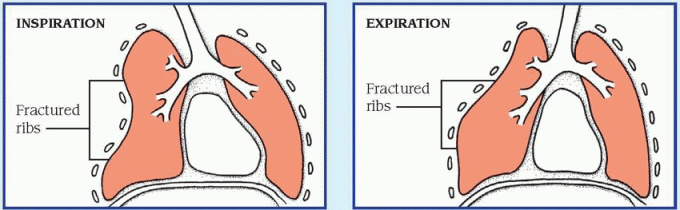
In flail chest—a disruption of the thorax due to multiple rib fractures—the unstable portion of the chest wall collapses inward during inspiration and balloons outward during expiration.
If you detect asymmetrical chest expansion, first consider traumatic injury to the patient’s ribs or sternum, which can cause flail chest, a lifethreatening emergency characterized by paradoxical chest movement. Quickly take the patient’s vital signs and look for signs of acute respiratory distress—rapid and shallow respirations, tachycardia, and cyanosis. Use tape or sandbags to temporarily splint the unstable flail segment.
Depending on the severity of respiratory distress, administer oxygen by nasal cannula, mask, or mechanical ventilator. Insert an I.V. catheter to allow fluid replacement and administration of pain medication. Draw a blood sample from the patient for arterial blood gas analysis, and connect the patient to a cardiac monitor.
Although asymmetrical chest expansion may result from hemothorax, tension pneumothorax, bronchial obstruction, and other life-threatening causes, it isn’t a cardinal sign of these disorders. Because any form of asymmetrical chest expansion can compromise the patient’s respiratory status, don’t leave the patient unattended, and be alert for signs of respiratory distress.
HISTORY AND PHYSICAL EXAMINATION
If you don’t suspect flail chest and if the patient isn’t experiencing acute respiratory distress, obtain a brief history. Asymmetrical chest expansion commonly results from mechanical airflow obstruction, so find out if the patient is experiencing dyspnea or pain during breathing. If so, does he feel short of breath constantly or intermittently? Does the pain worsen his feeling of breathlessness? Does repositioning, coughing, or any other activity relieve or worsen the patient’s dyspnea or pain? Is the pain more noticeable during inspiration or expiration? Can he inhale deeply?
Ask if the patient has a history of pulmonary or systemic illness, such as frequent upper respiratory tract infections, asthma, tuberculosis, pneumonia, or cancer. Has he had thoracic surgery? (This typically produces asymmetrical chest expansion on the affected side.) Also, ask about blunt or penetrating chest trauma, which may have caused pulmonary injury. Obtain an occupational history to find out if the patient may have inhaled toxic fumes or aspirated a toxic substance.
Next, perform a physical examination. Begin by gently palpating the trachea for midline positioning. (Deviation of the trachea usually indicates an acute problem requiring immediate intervention.) Then examine the posterior chest wall for areas of tenderness or deformity. To evaluate the extent of asymmetrical chest expansion, place your hands—fingers together and thumbs abducted toward the spine—flat on both sections of the lower posterior chest wall. Position your thumbs at the 10th rib, and grasp the lateral rib cage with your hands. As the patient inhales, note the uneven separation of your thumbs, and gauge the distance between them. Then repeat this technique on the upper posterior chest wall. Next, use the ulnar surface of your hand to palpate for vocal or tactile fremitus on both sides of the chest. To check for vocal fremitus, ask the patient to repeat “99” as you proceed. Note any asymmetrical vibrations and areas of enhanced, diminished, or absent fremitus. Then percuss and auscultate to detect air and fluid in the lungs and pleural spaces. Finally, auscultate all lung fields for normal and adventitious breath sounds. Examine the patient’s anterior chest wall, using the same assessment techniques.
MEDICAL CAUSES
♦ Bronchial obstruction. Life-threatening loss of airway patency may occur gradually or suddenly in bronchial obstruction. Typically, lack of chest movement indicates complete obstruction; chest lag signals partial obstruction. If air is trapped in the chest, you may detect intercostal bulging during expiration and hyperresonance on percussion. You may also note dyspnea, accessory muscle use, decreased or absent breath sounds, and suprasternal, substernal, or intercostal retractions.
♦ Flail chest. In this life-threatening injury to the ribs or sternum, the unstable portion of the chest wall collapses inward during inspiration and balloons outward during expiration (paradoxical movement). The patient may have ecchymoses, severe localized pain, or other signs of traumatic injury to the chest wall. He may also exhibit rapid, shallow respirations, tachycardia, and cyanosis.
♦ Hemothorax. Hemothorax is life-threatening bleeding into the pleural space that causes chest lag during inspiration. Other findings include signs of traumatic chest injury, stabbing pain at the injury site, anxiety, dullness on percussion, tachypnea, tachycardia, and hypoxemia. If hypovolemia occurs, you’ll note signs of shock, such as hypotension and rapid, weak pulse.
♦ Kyphoscoliosis. Abnormal curvature of the thoracic spine in the anteroposterior direction (kyphosis) and the lateral direction (scoliosis) gradually compresses one lung and distends the other. This produces decreased chest wall movement on the compressed-lung side and expands the intercostal muscles during inspiration on the opposite side. It can also produce ineffective coughing, dyspnea, back pain, and fatigue.
♦ Myasthenia gravis. Progressive loss of ventilatory muscle function produces asynchrony of the chest and abdomen during inspiration (“abdominal paradox”), which can lead to acute respiratory distress. Typically, the patient’s shallow respirations and increased muscle weakness cause severe dyspnea, tachypnea and, possibly, apnea.
♦ Phrenic nerve dysfunction. In this disorder, the paralyzed hemidiaphragm fails to contract downward, causing asynchrony of the thorax and upper abdomen on the affected side during inspiration (“abdominal paradox”). Its onset may be sudden, as in trauma, or gradual, as in infection or spinal cord disease. If the patient has underlying pulmonary dysfunction that contributes to hyperventilation, his inability to breathe deeply or to cough effectively may cause atelectasis of the affected lung.
♦ Pleural effusion. Chest lag at endinspiration occurs gradually in this life-threatening accumulation of fluid, blood, or pus in the pleural space. Usually, some combination of dyspnea, tachypnea, and tachycardia precedes chest lag; the patient may also have pleuritic pain that worsens with coughing or deep breathing. The area of the effusion is delineated by dullness on percussion and by egophony, bronchophony, whispered pectoriloquy, decreased or absent breath sounds, and decreased tactile fremitus. The patient may have a fever if infection caused the effusion.
♦ Pneumonia. Depending on whether fluid consolidation in the lungs develops unilaterally or bilaterally, asymmetrical chest expansion occurs as inspiratory chest lag or as chest-abdomen asynchrony. The patient typically has fever, chills, tachycardia, tachypnea, and dyspnea along with crackles, rhonchi, and chest pain that worsens during deep breathing. He may also be fatigued and anorexic and have a productive cough with rust-colored sputum.
♦ Pneumothorax. Entrapment of air in the pleural space can cause chest lag at endinspiration. This life-threatening condition also causes sudden, stabbing chest pain that may radiate to the arms, face, back, or abdomen and dyspnea unrelated to the chest pain’s severity. Other findings include tachypnea, decreased tactile fremitus, tympany on percussion, decreased or absent breath sounds over the trapped air, tachycardia, restlessness, and anxiety.
Tension pneumothorax produces the same signs and symptoms as pneumothorax, but they’re much more severe. A tension pneumothorax rapidly compresses the heart and great vessels, causing cyanosis, hypotension, tachycardia, restlessness, and anxiety. The patient may also develop subcutaneous crepitation of the upper trunk, neck, and face and mediastinal and tracheal deviation away from the affected side. Auscultation of a crunching sound over the precordium with each heartbeat indicates pneumomediastinum.
♦ Poliomyelitis. In this rare disorder, paralysis of the chest wall muscles and diaphragm produces chest-abdomen asynchrony (“abdominal paradox”), fever, muscle pain, and weakness. Other findings include decreased reflex response in the affected muscles and impaired swallowing and speaking.
♦ Pulmonary embolism. This acute, lifethreatening disorder causes chest lag; sudden, stabbing chest pain; and tachycardia. The patient usually has severe dyspnea, blood-tinged sputum, pleural friction rub, and acute anxiety.
OTHER CAUSES
♦ Treatments. Asymmetrical chest expansion can result from pneumonectomy and surgical removal of several ribs. Chest lag or the absence of chest movement may also result from intubation of a mainstem bronchus, a serious complication typically due to incorrect insertion of an endotracheal tube or movement of the tube while it’s in the trachea.
SPECIAL CONSIDERATIONS
If you’re caring for an intubated patient, regularly auscultate breath sounds in the lung peripheries to help detect a misplaced tube. If this occurs, prepare the patient for a chest X-ray to allow rapid repositioning of the tube. Because asymmetrical chest expansion increases the work of breathing, supplemental oxygen is usually given during acute events.
PEDIATRIC POINTERS
Children are at greater risk than adults for inadvertent intubation of a mainstem bronchus (especially the left bronchus). Their breath sounds are usually referred from one lung to the other because of the small size of the thoracic cage, so use chest wall expansion as an indicator of correct tube position in children. Children also develop asymmetrical chest expansion, paradoxical breathing, and retractions with acute respiratory illnesses, such as bronchiolitis, asthma, and croup.
Congenital abnormalities, such as cerebral palsy and diaphragmatic hernia, can also cause asymmetrical chest expansion. In cerebral palsy, asymmetrical facial muscles usually accompany chest-abdomen asynchrony. In a lifethreatening diaphragmatic hernia, asymmetrical expansion usually occurs on the left side of the chest.
GERIATRIC POINTERS
Asymmetrical chest expansion may be more difficult to determine in elderly patients because of the structural deformities associated with aging.
Chest pain
Chest pain usually results from disorders that affect thoracic or abdominal organs—the heart, pleurae, lungs, esophagus, rib cage, gallbladder, pancreas, or stomach. An important indicator of several acute and life-threatening cardiopulmonary and GI disorders, chest pain can also result from a musculoskeletal or hematologic disorder, anxiety, and drug therapy.
Chest pain may arise suddenly or gradually, and its cause may be difficult to ascertain initially. The pain may radiate to the arms, neck, jaw, or back. It may be steady or intermittent and mild or acute, and it may range in character from a sharp shooting sensation to a feeling of heaviness, fullness, or even indigestion. Chest pain may be provoked or aggravated by stress, anxiety, exertion, deep breathing, or eating certain foods.
Ask the patient when his chest pain began. Did it develop suddenly or gradually? Is it more severe or frequent now than when it first started? Does anything relieve the pain? Does anything aggravate it? Ask the patient about associated symptoms. Sudden, severe chest pain requires prompt evaluation and treatment because it may herald a life-threatening disorder. (See Managing severe chest pain, pages 142 and 143.)
HISTORY AND PHYSICAL EXAMINATION
If the chest pain isn’t severe, proceed with the history. Ask if the patient feels diffuse pain or can point to the painful area. Sometimes a patient won’t perceive the sensation he’s feeling as pain, so ask whether he has any discomfort radiating to his neck, jaw, arms, or back. If he does, ask him to describe it. Is it a dull, aching, pressurelike sensation? A sharp, stabbing, knifelike pain? Does he feel it on the surface or deep inside? Find out whether it’s constant or intermittent. If it’s intermittent, how long does it last? Ask if movement, exertion, breathing, position changes, or eating certain foods worsens or helps relieve the pain. Does anything in particular seem to bring it on?
Review the patient’s history for cardiac or pulmonary disease, chest trauma, intestinal disease, or sickle cell anemia. Find out which medications he’s taking, if any, and ask about recent dosage or schedule changes.
Take the patient’s vital signs, noting tachypnea, fever, tachycardia, oxygen saturation, paradoxical pulse, and hypertension or hypotension. Also, look for jugular vein distention and peripheral edema. Observe the patient’s breathing pattern, and inspect his chest for asymmetrical expansion. Auscultate his lungs for pleural friction rub, crackles, rhonchi, wheezing, and diminished or absent breath sounds. Next, auscultate for murmurs, clicks, gallops, and pericardial friction rub. Palpate for lifts, heaves, thrills, gallops, tactile fremitus, and abdominal masses or tenderness. (See Chest pain: Causes and associated findings, pages 144 to 147.)
MEDICAL CAUSES
♦ Angina pectoris. A patient with angina pectoris may experience a feeling of tightness or pressure in the chest that he describes as pain or a sensation of indigestion or expansion. The pain usually occurs in the retrosternal region over a palm-sized or larger area. It may radiate to the neck, jaw, and arms—classically, to the inner aspect of the left arm. Angina tends to begin gradually, build to its maximum, then slowly subside. Provoked by exertion, emotional stress, or a heavy meal, the pain typically lasts 2 to 10 minutes (usually no longer than 20 minutes). Associated findings include dyspnea, nausea, vomiting, tachycardia, dizziness, diaphoresis, belching, and palpitations. You may hear an atrial gallop (a fourth heart sound [S4]) or a murmur during an anginal episode.
In Prinzmetal’s angina, caused by vasospasm of coronary vessels, chest pain typically occurs when the patient is at rest—or it may awaken him. It may be accompanied by dyspnea, nausea, vomiting, dizziness, and palpitations. During an attack, you may hear an atrial gallop.
♦ Anthrax (inhalation). This acute infectious disease is caused by the gram-positive, sporeforming bacterium Bacillus anthracis. Although the disease most commonly occurs in wild and domestic grazing animals, such as cattle, sheep, and goats, the spores can live in the soil for many years. The disease can occur in humans exposed to infected animals, tissue from infected animals, or biological agents. Most natural cases occur in agricultural regions worldwide. Anthrax may occur in cutaneous, inhalation, or GI forms.
Inhalation anthrax is caused by inhalation of aerosolized spores. Initial flulike signs and symptoms include fever, chills, weakness, cough, and chest pain. The disease generally occurs in two stages with a period of recovery after the initial signs and symptoms. The second stage develops abruptly and causes rapid deterioration marked by fever, dyspnea, stridor, and hypotension; death generally results within 24 hours. Radiologic findings include mediastinitis and symmetrical mediastinal widening.
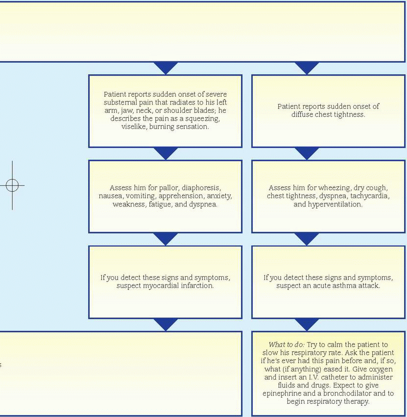
Managing severe chest pain
Sudden, severe chest pain may result from any one of several life-threatening disorders. Your evaluation and interventions will vary, depending on the pain’s location and character. The flowchart below will help you establish priorities for managing this emergency successfully.
Chest pain: Causes and associated findings
Major associated signs and symptoms
Common causes
Abdominal mass
Abdominal tenderness
Atrial gallop
Breath sounds, decreased
Cough
Crackles
Cyanosis
Diaphoresis
Dizziness
Dyspnea
Fever
Hemoptysis
Murmur
Nausea and vomiting
Pericardial friction rub
Pleural friction rub
Skin mottling
Syncope
Tachycardia
Tachypnea
Wheezing
Angina pectoris
•
•
•
•
•
•
•
Anthrax (inhalation)
•
•
•
Anxiety
•
•
•
•
•
Aortic aneurysm (dissecting)
•
•
•
•
•
•
•
Asthma
•
•
•
•
•
•
•
•
Blast lung injury
•
•
•
•
Blastomycosis
•
•
•
Bronchitis
•
•
•
•
•
Cardiomyopathy
•
•
•
•
•
•
Cholecystitis
•
•
•
•
•
Coccidioidomycosis
•
•
•
Costochondritis
•
Distention of colon’s splenic flexure
•
•
Esophageal spasm
•
•
Herpes zoster (shingles)
•
Hiatal hernia
•
Interstitial lung disease
•
•
•
•
Legionnaires’ disease
•
•
•
•
•
•
•
•
Lung abscess
•
•
•
•
•
•
•
•
Lung cancer
•
•
•
•
•
Mediastinitis
•
Mitral valve prolapse
•
•
•
•
Muscle strain
•
Myocardial infarction
•
•
•
•
•
•
•
Nocardiosis
•
•
•
•
Pancreatitis
•
•
•
•
•
•
Peptic ulcer
•
•
Pericarditis
•
•
•
•
Plague
•
•
•
•
•
Pleurisy
•
•
•
•
•
•
•
Pneumonia
•
•
•
•
•
•
•
•
Pneumothorax
•
•
•
•
•
Psittacosis
•
Pulmonary actinomycosis
•
•
•
Pulmonary embolism
•
•
•
•
•
•
•
•
•
•
•
Pulmonary hypertension (primary)
•
•
•
•
Q fever
•
Rib fracture
•
•
Sickle cell crisis
•
•
•
Thoracic outlet syndrome
•
Tuberculosis
•
•
•
•
Tularemia
•
•
•
♦ Anxiety. Acute anxiety—commonly known as panic attacks—can produce intermittent, sharp, stabbing pain, typically behind the left breast. This pain isn’t related to exertion and lasts only a few seconds, but the patient may experience a precordial ache or a sensation of heaviness that lasts for hours or days. Associated signs and symptoms include precordial tenderness, palpitations, fatigue, headache, insomnia, breathlessness, nausea, vomiting, diarrhea, and tremors. Panic attacks may be associated with agoraphobia—fear of leaving home or being in open places with other people.
♦ Aortic aneurysm (dissecting). The chest pain associated with this life-threatening disorder usually begins suddenly and is most severe at its onset. The patient describes an excruciating tearing, ripping, stabbing pain in his chest and neck that radiates to his upper back, abdomen, and lower back. He may also have abdominal tenderness, a palpable abdominal mass, tachycardia, murmurs, syncope, blindness, loss of consciousness, weakness or transient paralysis of the arms or legs, a systolic bruit, systemic hypotension, asymmetrical brachial pulses, lower blood pressure in the legs than in the arms, and weak or absent femoral or pedal pulses. His skin is pale, cool, diaphoretic, and mottled below the waist. Capillary refill time is increased in the toes, and palpation reveals decreased pulsation in one or both carotid arteries.
♦ Asthma. In a life-threatening asthma attack, diffuse and painful chest tightness arises suddenly along with a dry cough and mild wheezing, which progress to a productive cough, audible wheezing, and severe dyspnea. Related respiratory findings include rhonchi, crackles, prolonged expirations, intercostal and supraclavicular retractions on inspiration, accessory muscle use, flaring nostrils, and tachypnea. The patient may also experience anxiety, tachycardia, diaphoresis, flushing, and cyanosis.
♦ Blast lung injury. Caused by a percussive shock wave after an explosion, blast lung injury can cause severe chest pain and possibly tearing, contusion, edema, and hemorrhage of the lungs of affected people. Worldwide terrorist activity has recently increased the incidence of this condition, which may also cause dyspnea, hemoptysis, wheezing, and cyanosis. Chest Xrays, arterial blood gas measurements, and computed tomography scans are common diagnostic tools. Although no definitive guidelines exist for caring for those with blast lung injury, treatment is based on the nature of the explosion, the environment in which it occurred, and any chemical or biological agents involved.
♦ Blastomycosis. Besides pleuritic chest pain, this disorder initially produces signs and symptoms that mimic those of a viral upper respiratory tract infection: a dry, hacking, or productive cough (and sometimes hemoptysis), fever, chills, anorexia, weight loss, fatigue, night sweats, and malaise.
♦ Bronchitis. In its acute form, this disorder produces burning chest pain or a sensation of substernal tightness. It also produces a cough, initially dry but later productive, that worsens the chest pain. Other findings include a lowgrade fever, chills, sore throat, tachycardia, muscle and back pain, rhonchi, crackles, and wheezing. Severe bronchitis causes a fever of 101° to 102° F (38.3° to 38.9° C) and possibly bronchospasm with increased coughing and wheezing.
♦ Cardiomyopathy. In hypertrophic cardiomyopathy, angina-like chest pain may occur with dyspnea, a cough, dizziness, syncope, gallops, murmurs, and palpitations.
♦ Cholecystitis. This disorder typically produces abrupt epigastric or right-upperquadrant pain, which may be sharp or intensely aching. Steady or intermittent pain may radiate to the back or the right shoulder. Associated findings commonly include nausea, vomiting, fever, diaphoresis, and chills. Palpation of the right upper quadrant may reveal an abdominal mass, rigidity, distention, or tenderness. Murphy’s sign—inspiratory arrest elicited when the examiner palpates the right upper quadrant as the patient takes a deep breath— may also occur.
♦ Coccidioidomycosis. In this disorder, pleuritic chest pain occurs with a dry or slightly productive cough. Other effects include fever, rhonchi, wheezing, occasional chills, sore throat, backache, headache, malaise, marked weakness, anorexia, and a macular rash.
♦ Costochondritis. Pain and tenderness occur at the costochondral junctions, especially at the second costicartilage. The pain usually can be elicited by palpating the inflamed joint.
♦ Distention of colon’s splenic flexure. Central chest pain may radiate to the left arm in patients with this disorder. The pain may be relieved by defecation or the passage of flatus.
♦ Esophageal spasm. In this disorder, substernal chest pain may last up to an hour and may radiate to the neck, jaw, arms, or back. It commonly mimics the squeezing or dull sensation associated with angina. Other signs and symptoms include dysphagia for solid foods, bradycardia, and nodal rhythm.
♦ Herpes zoster (shingles). The pain of preeruptive herpes zoster may mimic that of myocardial infarction (MI). Initially, the pain is characteristically sharp, shooting, and unilateral. About 4 to 5 days after its onset, small, red, nodular lesions erupt on the painful areas— usually the thorax, arms, and legs—and the chest pain becomes burning. Associated findings include fever, malaise, pruritus, and paresthesia or hyperesthesia of the affected areas.
♦ Hiatal hernia. Typically, this disorder produces an angina-like sternal burning (heartburn), ache, or pressure that may radiate to the left shoulder and arm. The discomfort commonly occurs after a meal when the patient bends over or lies down. Other findings include a bitter taste and pain while eating or drinking, especially spicy foods and hot drinks.
♦ Interstitial lung disease. As this disease advances, the patient may experience pleuritic chest pain along with progressive dyspnea, cellophane-type crackles, a nonproductive cough, fatigue, weight loss, decreased exercise tolerance, clubbing, and cyanosis.
♦ Legionnaires’ disease. This disorder produces pleuritic chest pain in addition to malaise, headache, and possibly diarrhea, anorexia, diffuse myalgia, and general weakness. Within 12 to 24 hours, the patient suddenly develops a high fever and chills, and an initially nonproductive cough progresses to a productive cough with mucoid and then mucopurulent sputum and possibly hemoptysis. Patients may also experience flushed skin, mild diaphoresis, prostration, nausea and vomiting, mild temporary amnesia, confusion, dyspnea, crackles, tachypnea, and tachycardia.
♦ Lung abscess. Pleuritic chest pain develops insidiously in a lung abscess along with a pleural friction rub and a cough that produces copious amounts of purulent, foul-smelling, blood-tinged sputum. The affected side is dull on percussion, and decreased breath sounds and crackles may be heard. The patient also displays diaphoresis, anorexia, weight loss, fever, chills, fatigue, weakness, dyspnea, and clubbing.
♦ Lung cancer. The chest pain associated with lung cancer is commonly described as an intermittent aching felt deep within the chest. If the tumor metastasizes to the ribs or vertebrae, the pain becomes localized, continuous, and gnawing. Associated findings include a cough (sometimes blood-tinged), wheezing, dyspnea, fatigue, anorexia, weight loss, and fever.
♦ Mediastinitis. This disorder produces severe retrosternal chest pain that radiates to the epigastrium, back, or shoulder and may worsen with breathing, coughing, or sneezing. Accompanying signs and symptoms include chills, fever, and dysphagia.
♦ Mitral valve prolapse. Most patients with mitral valve prolapse are asymptomatic, but some may experience sharp, stabbing precordial chest pain or precordial ache. The pain can last for seconds or hours and may mimic the pain of ischemic heart disease. The characteristic sign of mitral prolapse is a midsystolic click followed by a systolic murmur at the apex. The patient may experience cardiac awareness, migraine headache, dizziness, weakness, episodic severe fatigue, dyspnea, tachycardia, mood swings, and palpitations.
♦ Muscle strain. Strained chest, arm, or shoulder muscles may cause a superficial and continuous ache or “pulling” sensation in the chest. Lifting, pulling, or pushing heavy objects may aggravate this discomfort. With acute muscle strain, the patient may experience fatigue, weakness, and rapid swelling of the affected area.
♦ Myocardial infarction. The crushing substernal chest pain typically associated with an MI lasts from 15 minutes to hours. Typically unrelieved by rest or nitroglycerin, the pain may radiate to the patient’s left arm, jaw, neck, or shoulder blades. Other findings include pallor, clammy skin, dyspnea, diaphoresis, nausea, vomiting, anxiety, restlessness, a feeling of impending doom, hypotension or hypertension, an atrial gallop, murmurs, and crackles.
An MI may be difficult to diagnose in perimenopausal women because it may produce atypical symptoms, such as fatigue, nausea, dyspnea, and shoulder or neck pain, rather than chest pain.
♦ Nocardiosis. This disorder causes pleuritic chest pain with a cough that produces thick, tenacious, purulent or mucopurulent, and possibly blood-tinged sputum. Nocardiosis may also cause fever, night sweats, anorexia, malaise, weight loss, and diminished or absent breath sounds.
♦ Pancreatitis. Acute pancreatitis usually causes intense epigastric pain that radiates to the back and worsens when the patient is in a supine position. Nausea, vomiting, fever, abdominal tenderness and rigidity, diminished bowel sounds, and crackles at the lung bases may also occur. A patient with severe pancreatitis may be extremely restless and have mottled skin, tachycardia, and cold, sweaty extremities. Fulminant pancreatitis causes massive hemorrhage, resulting in shock and coma.
♦ Peptic ulcer. In this disorder, sharp and burning pain usually arises in the epigastric region. This pain characteristically occurs hours after food intake, commonly during the night. It lasts longer than angina-like pain and is relieved by food or an antacid. Other findings include nausea, vomiting (sometimes with blood), melena, and epigastric tenderness.
♦ Pericarditis. This disorder produces precordial or retrosternal pain that’s aggravated by deep breathing, coughing, position changes, and occasionally by swallowing. The pain is commonly sharp or cutting and radiates to the shoulder and neck. Associated signs and symptoms include pericardial friction rub, fever, tachycardia, and dyspnea. Pericarditis usually follows a viral illness, but several other causes should be considered.
♦ Plague. Caused by Yersinia pestis, plague is one of the most virulent and, if untreated, most lethal bacterial infections known. Most cases are sporadic, but the potential for epidemic spread still exists. Clinical forms include bubonic (the most common), septicemic, and pneumonic plagues. The bubonic form is transmitted to man from the bite of infected fleas. Signs and symptoms include fever, chills, and swollen, inflamed, and tender lymph nodes near the site of the fleabite. Septicemic plague may develop as a complication of untreated bubonic or pneumonic plague and occurs when the plague bacteria enter the bloodstream and multiply. The pneumonic form can be contracted by inhaling respiratory droplets from an infected person or inhaling the organism that has been dispersed in the air through biological warfare. The onset is usually sudden with chills, fever, headache, and myalgia. Pulmonary signs and symptoms include a productive cough, chest pain, tachypnea, dyspnea, hemoptysis, increasing respiratory distress, and cardiopulmonary insufficiency.
♦ Pleurisy. The sharp, even knifelike chest pain of pleurisy arises abruptly and reaches maximum intensity within a few hours. The pain is usually unilateral and located in the lower and lateral aspects of the chest. Deep breathing, coughing, or thoracic movement characteristically aggravates it. Auscultation over the painful area may reveal decreased breath sounds, inspiratory crackles, and a pleural friction rub. Dyspnea, rapid and shallow breathing, cyanosis, fever, and fatigue may also occur.
♦ Pneumonia. This disorder produces pleuritic chest pain that increases with deep inspiration and is accompanied by shaking chills and fever. The patient has a dry cough that later becomes productive. Other signs and symptoms include crackles, rhonchi, tachycardia, tachypnea, myalgia, fatigue, headache, dyspnea, abdominal pain, anorexia, cyanosis, decreased breath sounds, and diaphoresis.
♦ Pneumothorax. Spontaneous pneumothorax, a life-threatening disorder, causes sudden severe, sharp chest pain that increases with chest movement; it’s typically unilateral and rarely localized. When the pain is centrally located and radiates to the neck, it may mimic that of an MI. After the pain’s onset, dyspnea and cyanosis progressively worsen. Breath sounds are decreased or absent on the affected side with hyperresonance or tympany, subcutaneous crepitation, and decreased vocal fremitus. Asymmetrical chest expansion, accessory muscle use, a nonproductive cough, tachypnea, tachycardia, anxiety, and restlessness also occur.
♦ Psittacosis. This disorder may produce pleuritic chest pain on rare occasions. It typically begins abruptly with chills, fever, headache, myalgia, epistaxis, and prostration.
♦ Pulmonary actinomycosis. This disorder causes pleuritic chest pain with a cough that’s initially dry but later produces purulent sputum. The patient may also display hemoptysis, fever, weight loss, fatigue, weakness, dyspnea, and night sweats. Multiple sinuses may extend through the chest wall and drain externally.
♦ Pulmonary embolism. This disorder produces chest pain or a choking sensation. Typically, the patient first experiences sudden dyspnea with intense angina-like or pleuritic pain aggravated by deep breathing and thoracic movement. Other findings include tachycardia, tachypnea, cough (nonproductive or producing blood-tinged sputum), low-grade fever, restlessness, diaphoresis, crackles, pleural friction rub, diffuse wheezing, dullness on percussion, signs of circulatory collapse (weak, rapid pulse; hypotension), paradoxical pulse, signs of cerebral ischemia (transient unconsciousness, coma, seizures), signs of hypoxia (restlessness) and, particularly in the elderly, hemiplegia and other focal neurologic deficits. Less-common signs include massive hemoptysis, chest splinting, and leg edema. A patient with a large embolus may have cyanosis and distended neck veins.
♦ Pulmonary hypertension (primary). Anginalike pain develops late in patients with this disorder, usually on exertion. The precordial pain may radiate to the neck but doesn’t characteristically radiate to the arms. Typical accompanying signs and symptoms include exertional dyspnea, fatigue, syncope, weakness, cough, and hemoptysis.
Only gold members can continue reading. Log In or Register to continue
 If you detect carpopedal spasm, quickly examine the patient for signs of respiratory distress (laryngospasm, stridor, loud crowing noises, cyanosis) or cardiac arrhythmias, which indicate hypocalcemia. Obtain blood samples for electrolyte analysis (especially calcium), and perform an electrocardiogram. Connect the patient to a monitor to watch for the appearance of arrhythmias. Administer an I.V. calcium preparation, and provide emergency respiratory and cardiac support. If a calcium infusion doesn’t control seizures, administer a sedative, such as chloral hydrate or phenobarbital.
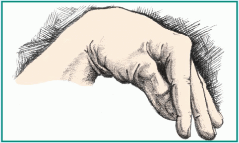
 Cri du chat syndrome is more common in females than males.
 Suspect cri du chat syndrome if you detect cat’s cry in a neonate. Be alert for signs of respiratory distress, such as nasal flaring; irregular, shallow respirations; cyanosis; and a respiratory rate over 60 breaths/minute. Be prepared to suction the neonate and to administer warmed oxygen. Keep emergency resuscitation equipment nearby because bradycardia may develop.
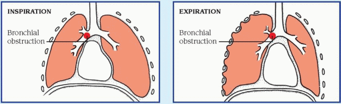
 If you detect asymmetrical chest expansion, first consider traumatic injury to the patient’s ribs or sternum, which can cause flail chest, a lifethreatening emergency characterized by paradoxical chest movement. Quickly take the patient’s vital signs and look for signs of acute respiratory distress—rapid and shallow respirations, tachycardia, and cyanosis. Use tape or sandbags to temporarily splint the unstable flail segment.
 Ask the patient when his chest pain began. Did it develop suddenly or gradually? Is it more severe or frequent now than when it first started? Does anything relieve the pain? Does anything aggravate it? Ask the patient about associated symptoms. Sudden, severe chest pain requires prompt evaluation and treatment because it may herald a life-threatening disorder. (See Managing severe chest pain, pages 142 and 143.)
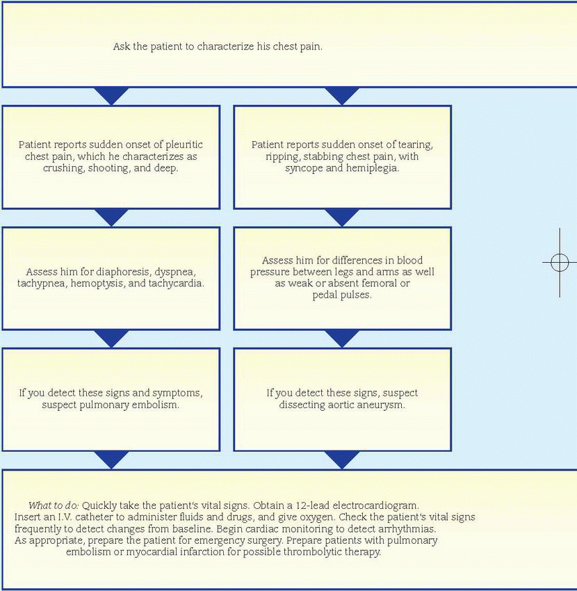
 An MI may be difficult to diagnose in perimenopausal women because it may produce atypical symptoms, such as


