Bronchiectasis
Alvaro C. Laga
Timothy C. Allen
Philip T. Cagle
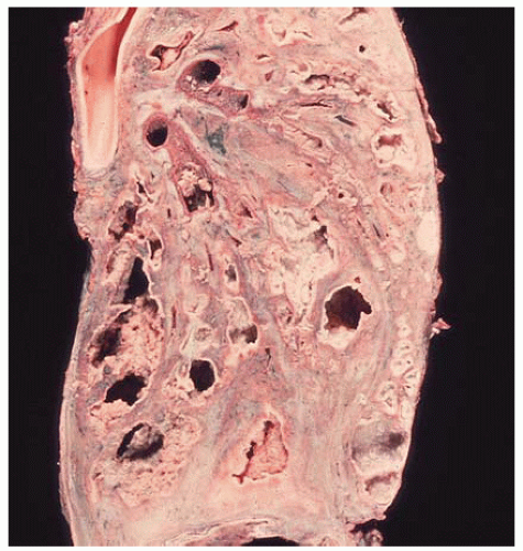
Bronchiectasis refers to the irreversible pathologic dilation of bronchi due to inflammation and fibrosis, most often associated with chronic infection. There are many underlying predisposing conditions, including airway obstruction (tumors, foreign bodies, mucous plugs), cystic fibrosis, Kartagener syndrome, and immune deficiency. The lower lobes are more commonly involved.
Histologic Features
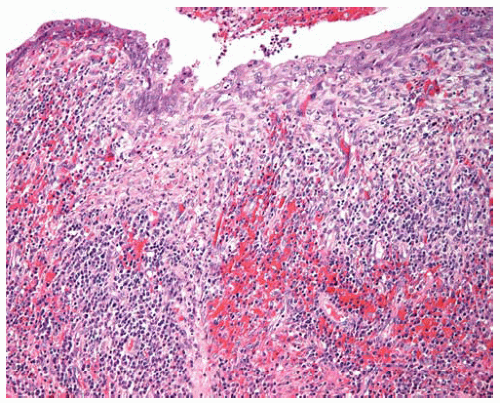
Varying degrees of inflammation, ulceration, and squamous metaplasia may be present within dilated bronchi.
Lymphoid follicles are frequently observed in the bronchial walls.
Extensive destruction of the bronchial wall including cartilage, muscle, and submucosal glands.
Fibrosis and inflammation of the bronchial wall and peribronchial tissues.
Associated acute and chronic pneumonias, organizing pneumonia, and postobstructive fibrosis and inflammation.
 Figure 57.1 Gross figure of bronchiectasis showing dilated infected segmental bronchi and fibrosis of surrounding lung parenchyma. |
 Figure 57.2 Bronchus with squamous metaplasia, mucosal erosion, granulation tissue, chronic inflammation, and fibrosis. |
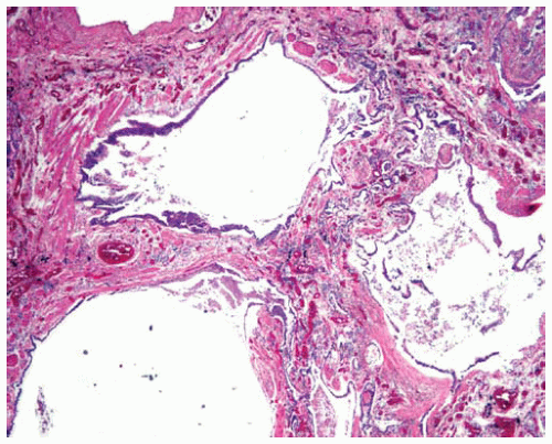
 Figure 57.3 Honeycomb lung due to bronchiectasis consists of residual spaces lined by metaplastic columnar epithelium within severe fibrosis with smooth muscle hyperplasia. |
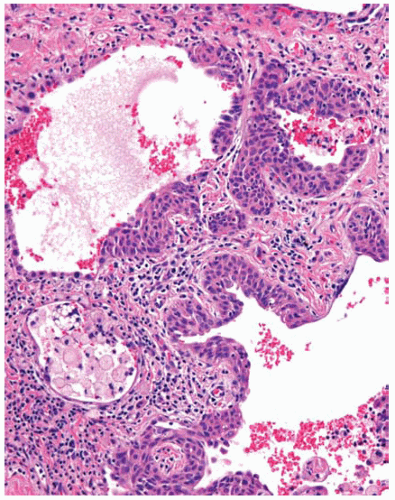
 Figure 57.4 Severe fibrosis due to bronchiectasis with residual spaces lined by metaplastic squamous epithelium with lipidladen macrophages within a residual space.
Stay updated, free articles. Join our Telegram channel
Full access? Get Clinical Tree
 Get Clinical Tree app for offline access

|